
Learn how the human eye captures, refracts, and processes light. Discover the mechanics of accommodation, retinal processing, and binocular vision.
The Physiology of Sight: How the Human Eye Captures and Processes Vision
Human eyesight is a sophisticated physiological process requiring perfect coordination between ocular structures and the brain. The eye acts as an organic camera, capturing ambient light rays, bending them precisely to a sharp focal point, and converting them into electrical data.
The Step-by-Step Pathway of Light
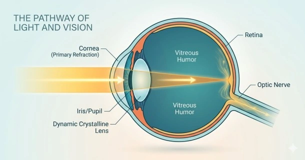
For the brain to interpret a clear image, light must travel unimpeded through a specific series of transparent anatomical structures:
[Ambient Light] ➔ Cornea ➔ Aqueous Humor ➔ Pupil/Iris ➔ Crystalline Lens ➔ Vitreous Humor ➔ Retina ➔ Optic Nerve ➔ Brain (Visual Cortex)- Refraction at the Cornea: Light reflects off external objects and hits the cornea. The cornea provides the majority of the eye's fixed focusing power.
- Passage through the Aqueous Humor: Light travels through this clear, nourishing fluid.
- Regulation at the Pupil: The iris contracts or dilates its central opening. This dynamically controls internal light exposure.
- Fine-Tuning at the Lens: The crystalline lens bends the light further.
- Focusing onto the Retina: Light spans across the vitreous cavity to strike the retina.
The Mechanism of Dynamic Accommodation
The human eye must constantly adjust to view objects at varying distances. This real-time focus correction is driven by a process called accommodation:
- Distant Tracking: Ciliary muscles relax, tightening the suspension fibers. This flattens the lens to focus distant light waves.
- Near Tracking: Ciliary muscles contract, releasing structural fiber tension. This thickens the lens, increasing its light-bending power.
Phototransduction: Converting Light into Brain Data
Once light waves hit the rear tissue of the retina, millions of photoreceptor cells initiate a biochemical process called phototransduction:
- Cone Cells: Concentrated within the central macula. They generate high-acuity central vision, sharp details, and full-color perception.
- Rod Cells: Located heavily across the peripheral retina. They operate under low illumination, tracking peripheral movement and enabling night vision.
These specialized cells instantly translate physical photon impacts into electrical neural signals.
Neurological Integration & Binocular Vision
True depth perception relies entirely on binocular vision. Because our eyes are physically separated on the face, each eye captures a slightly different angle of a target scene.
The two distinct streams of electrical impulses travel simultaneously along the optic nerves. They meet and cross at the optic chiasm before arriving at the brain's occipital lobe. The visual cortex seamlessly fuses these overlapping images into a single, three-dimensional panoramic view, allowing us to accurately calculate distance, speed, and depth.
Structural Variations: Why Eyes Go Out of Focus
When the physical shape of the eyeball or the curvature of the cornea deviates from ideal alignment, light fails to hit the retina perfectly, resulting in blurred vision:
- Myopia (Nearsightedness): The eyeball is too elongated, or the cornea is overly curved, causing light to focus in front of the retina.
- Hyperopia (Farsightedness): The eyeball is too short, causing light to reach a theoretical focus point behind the retina.
- Astigmatism: An irregular, football-shaped cornea distorts light across multiple focal points, blurring vision at all distances.
These anatomical variations require corrective prescription eyeglasses or contact lenses to re-route light path trajectories directly back onto the retinal plane.
Frequently Asked Questions (FAQ)
How does the human eye adapt so quickly when walking into a dark room?
This quick adaptation relies on two simultaneous processes. First, the iris muscles immediately dilate the pupil to let in maximum light. Second, your rod photoreceptors undergo a chemical reset, ramping up their sensitivity to navigate low-light environments safely.
Why do we lose the ability to read small print close-up as we get older?
This condition is called presbyopia. It occurs because the crystalline lens naturally hardens and loses its elasticity over time. As the lens stiffens, the ciliary muscles can no longer reshape it effectively to focus on near objects, requiring reading glasses.
What happens in the brain if the two eyes send completely mismatched images?
If the brain receives mismatched images due to eye misalignment, it can cause double vision. To prevent confusion, the brain may permanently suppress or ignore the weaker visual stream. If left untreated during childhood, this neurological suppression leads to a permanent drop in vision known as amblyopia.