
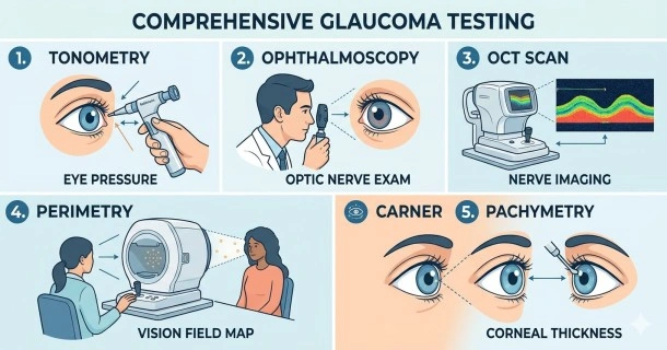
How is glaucoma diagnosed? Learn about tonometry, visual field tests, OCT nerve scans, and why eye pressure isn't the only metric that matters.
Glaucoma Testing: How is Glaucoma Diagnosed?
For decades, many believed that a simple eye pressure check was all it took to detect glaucoma. Today, clinical practice has evolved. A comprehensive glaucoma diagnosis relies heavily on evaluating the structural health of the optic nerve, rather than just measuring intraocular pressure (IOP).
Because variants like normal-tension glaucoma present with perfectly average eye pressure, relying on pressure alone leaves many at risk. Confirming a diagnosis requires tracking progressive changes in nerve tissue, mapping out blind spots, and assessing the physical anatomy of the eye's drainage systems.
The 5 Standard Diagnostic Glaucoma Tests
An eye care professional—an optometrist or ophthalmologist—will rarely rely on a single test. Instead, they administer a specialized suite of assessments to build a complete profile of your optical health.
1. Tonometry (Measuring Intraocular Pressure)
This test measures the fluid pressure inside your eye.
- How it works: The clinical gold standard is Goldmann applanation tonometry. After applying numbing drops, a small probe gently touches your cornea to measure its resistance. Alternative methods include hand-held pens or the "air puff" test.
- What it detects: While a normal pressure range is generally between 10 to 21 mm Hg, elevated pressure is a primary risk factor, though not a standalone confirmation of glaucoma.
2. Ophthalmoscopy & Imaging (Evaluating the Optic Nerve)
Because glaucoma destroys nerve fibers, examining the back of the eye is the most critical diagnostic step.
- How it works: Your pupils are dilated using specialized drops so the doctor can look directly at the shape, color, and structural integrity of the optic nerve.
- Advanced Scans: Clinics frequently use Optical Coherence Tomography (OCT) or digital fundus photography. These devices take high-resolution cross-sectional scans of your retina to measure the precise thickness of the retinal nerve fiber layer (RNFL), catching microscopic tissue loss long before you notice vision changes.
3. Perimetry (Visual Field Testing)
This mapping test measures your complete scope of vision, specifically identifying any early patches of peripheral vision loss.
- How it works: You sit in front of a dome-shaped machine, look straight ahead at a fixed target, and press a button every time you see a tiny flash of light appear in your side vision.
- What it detects: It reveals whether the brain is compensating for localized blind spots, mapping out the progression of any functional vision loss.
4. Gonioscopy (Inspecting the Drainage Angle)
This anatomical test determines whether your glaucoma is open-angle or closed-angle.
- How it works: After applying numbing drops, a specialized mirrored contact lens is placed briefly on the eye.
- What it detects: It allows the specialist to physically look at the drainage angle where the iris meets the cornea. This reveals whether the drain is wide open, narrowed, or entirely blocked by scar tissue or an overlapping iris.
5. Pachymetry (Measuring Corneal Thickness)
The thickness of your cornea directly influences how eye pressure readings are calculated.
- How it works: A tiny ultrasonic probe is lightly tapped against the front of the eye to measure corneal depth in micrometers.
- What it detects: A naturally thin cornea can artificially lower your pressure reading on a tonometer, masking dangerously high internal pressure. Individuals with thin corneas are statistically at a much higher risk for progressive nerve damage.
Tracking the "Glaucoma Suspect"
Sometimes, an initial exam reveals an atypical optic nerve shape or borderline high pressure, but without explicit tissue loss or visual field defects. These individuals are classified as glaucoma suspects. Because glaucoma is a chronic, progressive condition, suspects do not necessarily need immediate treatment, but they do require routine monitoring over time to catch early changes.
Glaucoma Test Overview
| Test Name | What It Measures | Why It Matters |
| Tonometry | Intraocular Pressure (IOP) | Identifies pressure spikes or abnormalities |
| Ophthalmoscopy / OCT | Optic Nerve Structure & RNFL Thickness | Direct evidence of structural nerve damage |
| Perimetry | Central & Peripheral Visual Fields | Detects functional blind spots or tunnel vision |
| Gonioscopy | The Iris-Cornea Drainage Angle | Differentiates open-angle from closed-angle |
| Pachymetry | Microscopic Corneal Thickness | Corrects and calibrates true pressure readings |
| Test Name | What It Measures | Why It Matters |
|---|---|---|
| Tonometry | Intraocular Pressure (IOP) | Identifies pressure spikes or abnormalities |
| Ophthalmoscopy / OCT | Optic Nerve Structure & RNFL Thickness | Direct evidence of structural nerve damage |
| Perimetry | Central & Peripheral Visual Fields | Detects functional blind spots or tunnel vision |
| Gonioscopy | The Iris-Cornea Drainage Angle | Differentiates open-angle from closed-angle |
| Pachymetry | Microscopic Corneal Thickness | Corrects and calibrates true pressure readings |
Frequently Asked Questions
Is tonometry painful?
No, modern tonometry testing is not painful. Before the procedure, the eye doctor administers quick-acting anesthetic drops that entirely numb the surface of your eye. You might feel a light pressure or the sensation of a cool mist, but you will not feel any pain.
How long do dilation eye drops last during a glaucoma test?
Dilation drops typically take 20 to 30 minutes to fully open the pupils and generally wear off within 4 to 6 hours. During this window, your eyes will be highly sensitive to bright light, and your near vision will be noticeably blurry. It is highly recommended to bring sunglasses and arrange for a driver to take you home.
Why do I need a visual field test if I can see perfectly?
Glaucoma damages the outer edges of your vision first. Because both of your eyes work together, your brain automatically fills in patchy blind spots, making your vision feel perfectly normal. A perimetry test isolates each eye to map out hidden blind spots before they spread to your central vision.