Wondering how a clinical nearsightedness diagnosis is made? Learn about visual acuity charts, phoropter testing, retinoscopy, and modern myopia tests.
Myopia Test: Comprehensive Guide to Nearsightedness Diagnosis
A clinical nearsightedness diagnosis is determined through a comprehensive eye examination performed by a licensed optometrist or ophthalmologist. Because progressive myopia can alter the physical shape of the eye, regular testing is critical for early intervention and overall vision preservation.
During a comprehensive myopia eye exam, eye care specialists use multiple clinical procedures to measure how your eyes focus light and determine the exact corrective lens power required.
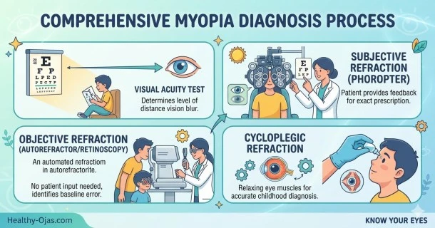
1. Visual Acuity Test (Snellen Eye Chart)
The foundational step in a myopia diagnosis test is evaluating visual acuity.
- The Procedure: Patients read rows of standardized letters or symbols from a Snellen chart placed at a specific distance (typically 20 feet or 6 meters).
- The Measurement: Visual acuity is recorded as a fraction (e.g., 20/40). The top number represents the standard testing distance (20 feet). The bottom number indicates the distance at which an eye with normal vision could read that same line. A recording of 20/40 means you must be 20 feet away to read a letter that a person with normal sight can read from 40 feet.
2. Subjective Refraction Testing (Phoropter)
To determine the precise prescription for eyeglasses or contact lenses, clinicians use an instrument called a phoropter.
- The Procedure: You sit comfortably behind the phoropter and look through different lens combinations at a distance chart.
- The Evaluation: The eye doctor flips through various lens powers, asking for your subjective feedback on which options yield the sharpest, clearest image. This interactive process pinpoints your exact refractive error.
3. Objective Refraction (Retinoscopy & Autorefraction)
Objective testing allows eye care professionals to measure refractive errors without relying entirely on patient feedback. This is particularly essential for a pediatric myopia evaluation where young children may not be able to articulate their vision changes verbally.
- Retinoscopy: The practitioner shines a specialized light from a retinoscope into the eye and observes the light reflection (reflex) off the retina. By moving the light back and forth across the pupil, the doctor calculates the eye's focusing power.
- Autorefraction: Modern clinics frequently utilize automated autorefractors, which instantly analyze how light changes as it enters the eye to provide an accurate baseline prescription.
4. Cycloplegic Refraction (Using Eye Drops)
In many cases—especially when evaluating children or managing severe eye fatigue—eye specialists perform a cycloplegic refraction.
- The Procedure: Formulated diagnostic eye drops are administered to temporarily relax the eye’s internal focusing muscles (the ciliary muscles).
- The Clinical Benefit: These drops prevent the eye from subconsciously over-focusing (accommodating) during the exam. This isolates the true structural refractive power of the eye and rules out temporary conditions like pseudo-myopia (false nearsightedness caused by muscle spasms).
3 Key Diagnostic Possibilities
When evaluating distance blur, clinicians perform these tests to rule out or identify distinct visual conditions:
- True Structural Myopia: Permanent blur caused by an elongated eyeball or an overly steep cornea.
- Fluctuating Blood Glucose: Rapid systemic blood sugar shifts can draw fluid into the crystalline lens, causing it to swell and induce temporary nearsightedness.
- Nuclear Sclerotic Cataracts: Early-stage age-related cataracts increase the density of the crystalline lens, creating a temporary "myopic shift" that degrades distance vision.
Myopia Treatment and Management Options
Once a clinical diagnosis is confirmed, eye care providers look beyond standard corrective lenses to offer comprehensive management plans aimed at slowing axial eye growth:
- Optical Defocus Lenses: Specialized eyeglasses or multifocal contact lenses designed to signal the eye to slow down its elongation rate.
- Orthokeratology (Ortho-K): Custom rigid, gas-permeable contact lenses worn overnight to temporarily reshape the cornea for clear daytime vision without glasses.
- Lifestyle Modifying Habits: Implementing the 20-20-20 rule during close-up tasks and increasing natural outdoor daylight exposure to 90–120 minutes daily to encourage healthy ocular development.
Always make sure to double-check the physical label and parameter packaging of your corrective lenses to ensure proper wear schedules and compliance.
Frequently Asked Questions (FAQ)
What should I expect during a comprehensive myopia eye exam?
A standard examination is completely painless. It involves reading letters from a distance chart (visual acuity testing), looking through a lens-switching device called a phoropter, and having the doctor examine the back of your eye using specialized lights and automated instruments.
Why does the eye doctor use eye drops for a child's myopia test?
Children have highly flexible focusing muscles inside their eyes. Diagnostic cycloplegic eye drops are used to temporarily relax these muscles so the doctor can measure the true physical shape and prescription of the eye without interference from muscle over-exertion.
Can a standard eye chart test fully diagnose myopia?
While a standard distance eye chart measures visual acuity and detects distance blur, it cannot determine the exact prescription or cause of the blur. Advanced tests using phoropters, autorefractors, and comprehensive ocular health checks are necessary for a definitive, accurate diagnosis.