
What causes retinal detachment? Discover the medical triggers behind rhegmatogenous, tractional, and exudative detachments, key risk factors, and myths.
Retinal Detachment Causes: Medical Triggers & Risk Factors Explained
Retinal detachment is a critical ophthalmic emergency that occurs when the light-sensitive layer of tissue at the back of the eye (the retina) pulls away from its underlying supportive layer of blood vessels. This separation deprives the retinal cells of oxygen and essential nutrients. If it is not treated promptly by an eye care specialist, it can lead to permanent, irreversible vision loss.
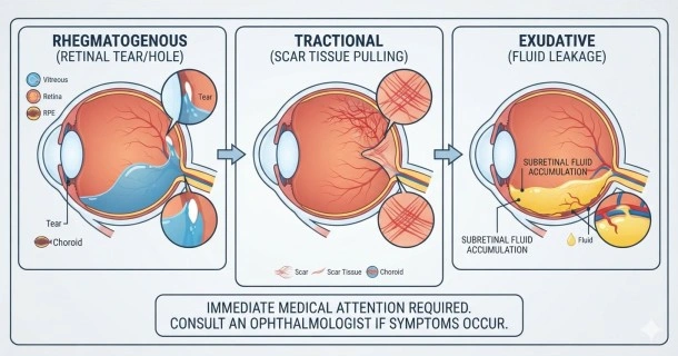
While many people search for a single cause, the medical community classifies retinal detachment causes into three distinct types depending on the underlying mechanism: rhegmatogenous, tractional, and exudative (serous).
The 3 Main Types of Retinal Detachment and Their Causes
1. Rhegmatogenous Retinal Detachment (The Most Common Cause)
A rhegmatogenous retinal detachment is the most frequent type diagnosed by ophthalmologists. It is directly caused by a full-thickness tear or hole in the retina.
- How it happens: When a tear forms, the liquid, gel-like vitreous humor that fills the center of your eye slips through the structural break. It begins to pool beneath the retina. As the fluid accumulates, the hydrostatic pressure progressively peels the retina away from the back of the eyeball.
- The Role of PVD: This process is most frequently triggered by a Posterior Vitreous Detachment (PVD). As a natural part of the aging process, the vitreous gel liquefies and shrinks. If the gel adheres too firmly to the retina as it separates, it can pull hard enough to rip the tissue, leaving behind a hazardous tear.
2. Tractional Retinal Detachment
A tractional retinal detachment develops without any initial tears or holes in the retinal tissue. Instead, it is caused by mechanical forces pulling from within the eye.
- How it happens: Advanced cellular scar tissue or abnormal fibrovascular membranes grow across the surface of the retina. As this scar tissue contracts, it exerts a physical "tugging" or traction force that physically lifts the retina out of its proper position.
- Primary Triggers: This type is a notorious complication of proliferative diabetic retinopathy. Severe, long-term uncontrolled diabetes damages the delicate blood vessels of the eye, causing them to leak and sprout fragile new vessels that scar over time. Other causes include sickle cell retinopathy, advanced ocular trauma, or previous intraocular surgical complications.
3. Exidative (Serous) Retinal Detachment
An exudative retinal detachment occurs completely independently of retinal tears or mechanical traction.
- How it happens: Fluid leaks directly out of the blood vessels situated beneath the retina (in the choroid bed) and collects in the subretinal space. The pooling fluid creates a physical barrier that pushes the retina forward, detaching it.
- Primary Triggers: This type is typically caused by severe inflammatory diseases (such as uveitis or sarcoidosis), eye infections (like cytomegalovirus retinitis), eye tumors (such as choroidal melanoma), or systemic vascular conditions like malignant hypertension.
Major Risk Factors: Who is Most Susceptible?
While anyone can experience a retinal detachment, clinical data shows you are at a significantly higher risk if you possess any of the following clinical indicators:
- Severe Nearsightedness (High Myopia): Individuals requiring strong corrective lenses for distance vision often have physically elongated eyeballs. This elongation stretches and thins the peripheral retina, making it highly susceptible to spontaneous holes, structural thinnings like lattice degeneration, and tears.
- Advanced Age: The risk rises sharply after the age of 40 to 50, primarily because the vitreous gel naturally shifts, liquefies, and contracts during these decades.
- Previous Eye Trauma: A blunt force blow or penetrating injury to the eye can cause immediate or delayed retinal dialyses (peripheral tears), even years after the incident occurred.
- Prior Intraocular Surgery: Procedures such as cataract extraction or glaucoma operations can destabilize the internal dynamics of the eye and induce premature posterior vitreous detachment.
- Family or Personal History: If you have experienced a detachment in one eye, you have an elevated risk of developing it in your fellow eye. A genetic family history of retinal diseases also increases vulnerability.
Debunking Common Myths: What Does Not Cause It?
There is significant misinformation online regarding everyday activities and retinal health. To prevent unnecessary panic, it is important to clarify what clinical studies have proven does not cause a detached retina:
- Heavy Lifting or Physical Strain: Standard exercise, heavy lifting, or intense physical exertion do not generate intraocular forces capable of detaching a healthy retina.
- Severe Eye Strain: Reading in low light, looking at digital screens for prolonged periods, or experiencing standard visual fatigue may cause temporary discomfort, dry eyes, or headaches, but it has no physical mechanism to cause retinal breaks or detachments.
Early Warning Signs: When to Seek Immediate Care
Retinal detachment is entirely painless, but it presents with distinct, undeniable visual disturbances. If you experience any of the following symptoms, you must visit an eye care professional or an emergency room immediately:
- A sudden, dramatic increase in visual floaters (specks, cobwebs, or threads drifting across your field of vision).
- Frequent, sudden flashes of light (photopsia) in one or both eyes, which are highly visible even in dark rooms.
- A dark shadow or curtain-like obstruction creeping in from your peripheral (side) vision.
- Sudden, unexplained blurring or distortion of your central vision.
Frequently Asked Questions (FAQ)
Can stress or high blood pressure cause retinal detachment?
Psychological stress does not directly cause the condition. However, severely elevated blood pressure (malignant hypertension) can damage the blood vessels behind the retina, leading to fluid leakage and a subsequent exudative (serous) retinal detachment.
How quickly does a retinal tear turn into a full detachment?
The progression timeline is highly unpredictable. A retinal tear can progress to a full detachment within a matter of hours, days, or it may take weeks or even months. Because there is no way to predict the speed of progression, any diagnosed retinal tear is treated as an urgent medical priority.
Can a detached retina fix itself over time?
No. A detached retina cannot heal or reattach on its own. It requires specialized surgical intervention—such as a vitrectomy, scleral buckling, or pneumatic retinopexy—to seal the tears and reposition the tissue securely against the back of the eye.