
Explore surgical treatments for a detached retina, including scleral buckling, vitrectomy, and pneumatic retinopexy. Learn about recovery timelines and success rates.
Retinal Detachment Surgery: Treatment Options & Recovery Guide
Retinal detachment surgery is the only effective treatment method available to repair a detached retina and prevent permanent blindness. Because retinal cells quickly die when separated from their blood supply, this condition is treated as an ophthalmologic emergency. Immediate surgical intervention is required to preserve your eyesight.
Depending on the severity, location, and specific type of your detachment, a vitreoretinal specialist will choose between outpatient preventative treatments or major intraocular surgical procedures.
Early Interventions: Treating Retinal Tears Before Detachment
If your ophthalmologist diagnoses a retinal tear or hole early, before fluid has accumulated underneath the tissue, non-invasive outpatient procedures can secure the area and prevent a full detachment from ever developing.
Laser Photocoagulation (Laser Sealing)
- How it works: A specialized medical laser beam is directed into the eye through a contact lens.
- The clinical mechanism: The laser creates microscopic, controlled burns around the perimeter of the retinal tear. As these tiny burns heal, they form a permanent chorioretinal scar that welds the retina back to its underlying wall, effectively blocking fluid from seeping underneath.
Retinal Cryopexy (Freezing Therapy)
- How it works: This method achieves the exact same goal as a laser but uses extreme cold instead of light energy.
- The clinical mechanism: A specialized freezing probe is applied to the external wall of the eye directly over the tear site. The intense cold creates a localized freeze-thaw reaction that induces a protective sealing scar. It is frequently utilized for tears located at the extreme peripheral edges of the retina that are physically difficult for lasers to reach.
Major Surgical Procedures for a Detached Retina
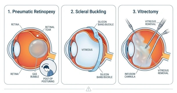
Once the retina has fully detached from the back wall of the eye, non-invasive lasers are no longer sufficient. Your surgeon will recommend one of three primary surgical techniques:
| Procedure Name | How It Reattaches the Retina | Typical Outpatient Context |
|---|---|---|
| Pneumatic Retinopexy | An expandable gas bubble is injected internally to push the retina flat against the eye wall. | Best for simple, single tears located in the upper half of the eye. |
| Scleral Buckling | A flexible silicone band is stitched to the outside of the eye, squeezing the eye wall inward. | Best for rhegmatogenous detachments without complex internal scar tissue. |
| Vitrectomy | The vitreous gel is fully removed and replaced with gas or temporary silicone oil. | Best for complex, tractional, or large-scale detachments. |
1. Pneumatic Retinopexy
During this office-based procedure, the surgeon treats the tear with cryopexy and then injects a tiny bubble of expandable gas into the vitreous cavity.
- The Mechanism: The bubble floats to the top of the eye. By precisely positioning your head, the bubble presses against the tear, squeezing out the trapped subretinal fluid.
- Patient Compliance: The gas bubble is gradually reabsorbed by the body over several weeks. However, success depends heavily on the patient maintaining a highly specific head position for several days post-op.
2. Scleral Buckling
This is a traditional, highly effective surgical technique performed in an operating room.
- The Mechanism: The surgeon sutures a fine band of silicone rubber or sponge onto the sclera (the white outer wall of the eye). This creates an indentation or "buckle" that pushes the eye wall inward to meet the detached retina.
- Long-Term Status: The silicone band is permanently left on the eye. It is completely invisible on the outside and cannot be felt by the patient after healing.
3. Vitrectomy
A vitrectomy is an advanced microsurgical procedure performed under local or general anesthesia.
- The Mechanism: The surgeon creates microscopic incisions to remove the vitreous gel from the center of the eye. This instantly relieves any mechanical traction pulling on the retina.
- Internal Tamponade: The internal cavity is then filled with either a long-lasting gas bubble or medical-grade silicone oil to hold the retina flat. If silicone oil is used, a minor secondary procedure is required months later to remove it.
Understanding the Risks and Potential Complications
While retinal reattachment surgery has a high clinical success rate, all intraocular surgeries carry some inherent medical risks. Potential complications include:
- Accelerated Cataract Development: The presence of intraocular gas or oil frequently causes the eye's natural lens to cloud over within a year.
- Secondary Glaucoma: Post-operative inflammation or gas expansion can cause a temporary spike in intraocular pressure.
- Proliferative Vitreoretinopathy (PVR): This is the growth of new aggressive scar tissue after surgery. It can pull the retina back off the wall, requiring a repeat operation.
- Ocular Infection (Endophthalmitis): A rare but severe complication that requires immediate emergency antibiotic treatment.
Recovery Timeline, Post-Op Care, and Visual Outlook
Recovering from a retinal operation requires patience and strict adherence to your surgeon’s medical instructions.
- The First 48 Hours: Expect temporary bruising, mild aching, and minor swelling around the eyelids. Your visual acuity will be extremely blurry, which is completely normal.
- Medication Protocol: You will be prescribed a strict regimen of antibiotic and anti-inflammatory eye drops for 2 to 6 weeks to prevent infection and manage swelling.
- The Golden Rule of Gas Bubbles: If a gas bubble was used, you must never travel by air or undergo general anesthesia using nitrous oxide. The atmospheric pressure changes will cause the bubble to expand rapidly, causing blindness.
- Visual Outlook: Final visual recovery takes 2 to 6 months. Your ultimate visual clarity depends heavily on whether your central macula was detached, and how long the tissue remained separated before surgery.
Frequently Asked Questions (FAQ)
What is the overall success rate of retinal detachment surgery?
Approximately 85% to 90% of retinal detachment surgeries are successful on the first attempt. However, if complex scar tissue (PVR) develops during healing, a small percentage of patients will require a second or third corrective procedure to fully secure the retina.
How long does it take for vision to return after surgery?
Your vision will remain highly blurry for the first few weeks, especially if a gas bubble or silicone oil is inside the eye. Significant visual improvement typically begins once the gas bubble dissipates (around 2 to 8 weeks), with final visual stabilizing occurring between 3 to 6 months post-surgery.
How should I sleep after retinal detachment surgery?
If your surgeon utilized an internal gas bubble, you will be instructed to maintain a specific face-down or side-lying position for 7 to 14 days, even while sleeping. This precise positioning ensures the floating bubble continually presses against the retinal tear to keep it sealed.