
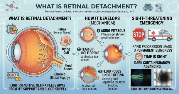
Curious about retinal health? Discover what retinal detachment is, how fluid shifts create tissue tears, and why it requires immediate medical intervention.
Understanding Retinal Detachment: An Essential Overview
Retinal detachment is a critical, sight-threatening medical emergency that occurs when the thin, light-sensitive nerve tissue layer at the back of the eye peels away from its underlying support structures and blood supply. If left untreated, the detachment can spread across the entire tissue layer, rapidly leading to permanent vision loss or complete blindness.
Because timely clinical intervention is the single most important factor in saving a patient's sight, recognizing what this condition is and understanding how it develops is vital for anyone at risk.
The Crucial Function of Your Retina
The retina functions much like the digital sensor or film inside a camera. When light enters the eye, the natural lens focuses it directly onto this delicate tissue network. The retina then instantly converts those captured light patterns into highly complex neural impulses, transmitting them through the optic nerve straight to the brain to be translated into the images you see.
When a detachment physically lifts the retina out of its proper anatomical position, it strips the photoreceptor cells of their vital oxygen and nutrient supply, causing them to immediately cease functioning.
How Retinal Detachment Develops
While there are multiple clinical mechanisms behind this disorder, the most frequent path begins with a microscopic break or tear in the retinal sheet.
- The Tearing Mechanism: As the eye ages, the gel-like vitreous fluid filling the center of the eyeball naturally shrinks and shifts. If the vitreous gel pulls too forcefully on the retina during this shift, it can cause a physical tear or hole to open.
- Fluid Accumulation: Once a break occurs, liquid vitreous fluid seeps through the hole and pools underneath the retina.
- The Separation: As the liquid continues to build up, the hydrostatic pressure gently lifts and separates the retina from the choroid layer underneath, blinding the affected section of your visual field.
Statistical Prevalence and Demographics
Retinal detachment can strike individuals at any point in their life, but it becomes markedly more common with advanced age.
- General Population: The baseline risk across the broad global population sits at approximately 5 in 100,000 individuals per year.
- Aging Demographics: The incidence rate spikes significantly among middle-aged and elderly populations, rising to roughly 20 in 100,000 individuals per year.
This entry serves as an introductory overview. To learn how to identify acute warning signs, explore what triggers the tissue split, or understand modern correction options, read our specialized chapters covering Retinal Detachment Symptoms, Causes and Risk Factors, and Emergency Repair Surgery Options.
Frequently Asked Questions (FAQ)
Can a detached retina heal on its own without emergency medical treatment?
No, a structural retinal detachment will not reattach or heal on its own. If left unaddressed by an eye specialist, the vitreous fluid will continuously seep under the tissue layer, expanding the separation until the entire retina detaches, which results in permanent legal blindness.
Who is most susceptible to developing a retinal detachment?
While age is a major driver, individuals with extreme nearsightedness (high myopia), a personal history of severe eye trauma, a family history of retinal tears, or those who have previously undergone cataract surgery carry a significantly higher clinical risk.
Is retinal detachment painful when it occurs?
No, retinal detachment is entirely painless because the light-sensitive tissue sheet lacks pain-sensing nerve fibers. Because you cannot feel it happening, you must rely entirely on visual warning indicators—such as sudden flashes of light, a flurry of dark spots (floaters), or a dark curtain shadow creeping across your vision—to know when to seek emergency help.