
Discover retinitis pigmentosa (RP), a rare inherited eye condition causing progressive rod-cone dystrophy. Learn about its timeline, genetic basis, and diagnostic baselines.
Retinitis Pigmentosa (RP): An Introduction to Progressive Retinal Degeneration
Retinitis Pigmentosa (RP) is not a single disease, but a complex group of rare, inherited genetic disorders that cause progressive degeneration of the retina. The retina is the light-sensitive tissue lining the back of the eye responsible for converting light into electrical signals for the brain.
As a classic form of rod-cone dystrophy, RP primarily targets the photoreceptor cells within the retina. This cellular breakdown typically leads to a predictable, gradual loss of vision over several decades. Because it is a progressive condition, early clinical awareness, comprehensive genetic testing, and low-vision management are vital to helping individuals navigate changes in their visual field.
Understanding the Core Disease Mechanism: Rods vs. Cones
To understand how retinitis pigmentosa compromises vision, it helps to understand the two primary types of photoreceptor cells located in the human retina:
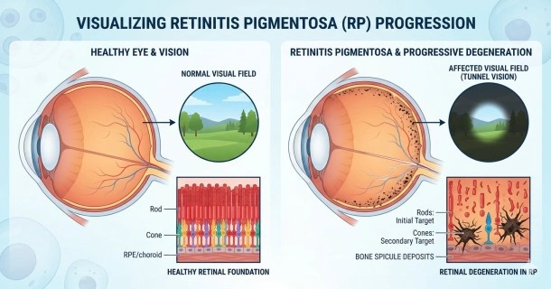
- Rod Photoreceptors (Primary Target): Rods are densely packed around the peripheral (outer) edges of the retina. They are responsible for your vision in dim light, night vision, and detecting motion in your side vision. In most forms of typical RP, rod cells degenerate first, which explains why night blindness is almost always the earliest clinical sign.
- Cone Photoreceptors (Secondary Target): Cones are concentrated in the central part of the retina (the macula). They are responsible for sharp central vision, fine details, and color perception. As RP advances, the progressive cellular damage eventually moves inward, compromising the cone cells and affecting central clarity.
The Typical Progression of Retinitis Pigmentosa
While the speed of vision loss varies widely depending on the specific genetic mutation involved, the clinical progression of retinitis pigmentosa generally follows a distinct timeline:
- Early Childhood / Adolescence: The first noticeable symptom is typically nyctalopia (severe night blindness). Affected individuals find it exceptionally difficult to adjust to dim environments, such as movie theaters or walking outdoors after sunset.
- Early Adulthood: The progressive loss of peripheral photoreceptors begins to constrict the visual field. This creates blind spots in the side vision, causing individuals to frequently bump into furniture or miss objects coming from the side.
- Advanced Stages: The visual field continues to shrink until only a small island of central vision remains. This clinical state is widely known as tunnel vision. While central visual acuity can remain intact for many years, advanced stages can eventually impact central clarity.
- For a comprehensive breakdown of early warning signs, visit our dedicated guide on Retinitis Pigmentosa Symptoms.
What Triggers This Condition? (Overview of Causes)
Retinitis pigmentosa is strictly an inherited genetic disorder. It is caused by mutations in any one of more than 100 different genes responsible for producing the proteins required for normal retinal function and survival.
These mutated genes are passed down through families via three primary patterns of inheritance: autosomal dominant, autosomal recessive, or X-linked. Notably, a lack of known family history does not rule out the condition. In fact, roughly 40% to 50% of diagnosed RP cases are classified as "simplex"—meaning the individual is the first in their family to manifest the condition, often due to hidden recessive genes or spontaneous new genetic mutations.
- To explore the inheritance patterns and genetic mutations in depth, read our full article on Retinitis Pigmentosa Causes.
How Do Specialists Spot and Diagnose RP?
An accurate, early diagnosis is essential to rule out mimicking retinal conditions and to establish a baseline for low-vision care. A retinal specialist or ophthalmologist will utilize a suite of advanced diagnostic tests:
- Electroretinography (ERG): Considered the gold standard for RP diagnosis, an ERG measures the collective electrical response of the retina's rods and cones to light stimuli. In patients with RP, the electrical readings are distinctly reduced or virtually unrecordable.
- Fundus Examination (Ophthalmoscopy): When looking inside the eye through a dilated pupil, a specialist can see hallmark physical signs of the disease. A classic RP retina displays dark, spiderweb-like patches of pigment called "bone spicule" pigment deposits, along with narrowed (attenuated) retinal blood vessels and a pale optic nerve.
- Visual Field Testing (Perimetry): This mapping test charts the full scope of your peripheral vision, precisely measuring the location and size of any developing blind spots.
- Learn exactly what to expect during your clinical evaluation in our guide to Retinitis Pigmentosa Diagnosis and Tests.
Are There Treatments Available?
Currently, there is no universal cure for retinitis pigmentosa, but the landscape of ophthalmic medicine is changing rapidly. Modern management focuses heavily on slowing disease progression, preserving remaining photoreceptors, and maximizing functional independence.
Management strategies include specialized low-vision aids, high-dose vitamin therapies under strict medical supervision, and cutting-edge gene therapies (such as Luxturna) designed for individuals with specific genetic mutations (like the RPE65 gene).
- To review the latest clinical options, emerging clinical trials, and adaptive tools, visit complete resource on Retinitis Pigmentosa Treatments.
Frequently Asked Questions (FAQ)
Does retinitis pigmentosa lead to total, absolute blindness?
While RP causes severe, progressive vision loss and results in legal blindness for many individuals, total absolute blindness (the inability to perceive light) is rare. Most individuals retain a small, functional window of central vision or light perception well into their later stages of life.
At what age do symptoms of retinitis pigmentosa typically begin?
The onset of symptoms most frequently occurs between childhood and the teenage years. However, because RP encompasses many different genetic mutations, some variations do not present noticeable vision changes until mid-adulthood.
Can you prevent retinitis pigmentosa if it runs in your family?
Because RP is an inherited genetic condition encoded in a person's DNA from birth, there is currently no way to prevent the development of the disorder. However, genetic counseling is highly recommended for families with a history of RP to help assess the risk of passing the mutated genes to future generations.