
Good Healthy Fats

Fats from nature are always good and healthy including saturated and natural trans-fat conjugated linoleic acid CLA. However it is healthy only if it is in moderation.
Trans-fats are Good! If it is from NATURE
Trans-fat conjugated linoleic acid or CLA is right for your health because naturally made in the bodies of ruminants (cows). The best sources of CLA are grass-fed beef (not factory-farmed, grain-fed) and raw dairy products from grass-fed cattle. CLA is useful for cancer, asthma, cardiovascular disease, hypertension, high cholesterol & triglycerides, osteoporosis, insulin resistance, inflammation, and obesity.
Benefits of Saturated Fats – Good fats
"The more saturated fat one ate, the more cholesterol one ate, the lower the person's serum (blood) cholesterol." -- William Castelli M.D., Framingham Director
Saturated fats are predominantly from animal products such as meat, dairy, and eggs. It is also in some plant sources such as coconut, palm, and palm kernel oils.
The myth that saturated fat causes heart disease has undoubtedly harmed a limitless number of lives over the past several decades.
Pacific Island populations get 30-60% of their total caloric from saturated oil (coconut oil). Nevertheless, there is non-existence of cardiovascular disease.
Association of Dietary, Circulating, and Supplement Fatty Acids with Coronary Risk: A Systematic Review and Meta-analysis published in Annals of Internal Medicine, 18 March 2014, Vol. 160, No. 6. This study shows the current evidence does not support cardiovascular guidelines that encourage high consumption of polyunsaturated fatty acids and low consumption of total saturated fats.
The cardiometabolic consequences of replacing saturated fats with carbohydrates or Ω-6 polyunsaturated fats: Do the dietary guidelines have it wrong? Open Heart 2014; 1: Date of issue: 10.1136/openhrt-2013-000032. The study shows the increase in the prevalence of diabetes and obesity in the USA occurred with an increase in the consumption of carbohydrate, not saturated fat. The benefits of a low-fat diet (particularly a diet replacing saturated fats with carbohydrates or Ω-6 polyunsaturated fatty acids) severely challenged. Dietary guidelines should assess the totality of the evidence and strongly reconsider their recommendations for replacing saturated fats with carbohydrates or Ω-6 polyunsaturated fats.
Importance of Saturated fat
The much-maligned saturated fats, which Americans are trying to avoid, are not the cause of our modern diseases. They play many essential roles in the body:
- Cell structure contains at least 50% saturated fatty acids. They give the cells necessary stiffness and integrity.
- Bones require saturated fat for their optimal health. The body needs at least 50% of the dietary fats from saturated fatty acid for the calcium effectively incorporating into the skeletal structure.
- Lp (a) is a substance in the blood prone to heart disease; saturated fat helps lower it. Ref: Plasma Lipoprotein (a) Levels in Men and Women Consuming Diets Enriched in Saturated, Cis, or Trans-Monounsaturated Fatty Acids, published in Arteriosclerosis, Thrombosis, and Vascular Biology, 1997; 17: 1657-1661. The study shows saturated fatty acids consistently decrease Lp(a) concentrations.
- Liver protection is by saturated fatty acids from the toxic insults of alcohol, other toxins & medications. It even can reverse the damage. Reference Dietary saturated fatty acids: a novel treatment for alcoholic liver disease published in Gastroenterology, 1995 Aug; 109(2):547-54. The study shows a diet enriched in saturated fatty acids reversed alcohol liver injury. Down-regulation of lipid peroxidation may explain this effect.
- Essential fatty acids such as omega-3 fatty acids are better remaining in the tissues when the diet is rich in saturated fats.
- Lung's airspaces are coating with a thin layer of lung surfactant made of 100 % saturated fatty acids. Replacing this fat by other fat types makes faulty surfactant causing breathing difficulties, airspace collapse, and respiratory problems.
- The brain mainly made of fat & cholesterol. Although unsaturated essential fatty acids in cold-water fish (EPA & DHA) are necessary for brain & nerve function, most of the fatty acids in the brain are saturated. The brain requires saturated fats for its optimal functioning.
- Nerve signaling is by certain saturated fats found in butter, lard, coconut, and palm oil. It functions as signaling messengers for hormone production, including insulin.
- The immune system depends on medium-chain saturated fatty acids such as myristic & lauric found in butter and coconut oil, which have potent germ-killing ability. Loss of these fatty acids in the white blood cells hampers their ability to recognize and destroy foreign invaders, such as viruses, bacteria, and fungi.
- Heart muscle cells prefer saturated long-chain palmitic and stearic acid to carbohydrates.
It is not surprising that saturated fats make up 54 % of the fat in the mother’s breast milk.
Saturated fat and coronary heart disease
A study by D.M. Dreon et al., titled “Change in Dietary Saturated Fat Intake Correlated with Change in Mass of Large Low-density Lipoprotein Particles in Men,” published in American Journal of Clinical Nutrition 67, no. 5 (1998): 828–36. This study shows eating fewer saturated fats may result in lower cholesterol level. However, the reduction is only large velvety good LDL make up a pattern A cholesterol profile. Conversely, if there is increasing saturated fat intake and decreasing carbohydrate intake, a significant shift to more prominent, fluffy, harmless LDL particles and makes up a pattern B cholesterol profile.
A study "Dietary fats, carbohydrate, and progression of coronary atherosclerosis in postmenopausal women" was published in The American Journal of Clinical Nutrition November 2004 volume 80 number 5 1175-1184. The study shows, with relatively low total fat intake, a higher saturated fat intake associated with less progression of atherosclerosis, whereas carbohydrate intake associated with more significant progress.
A study "Saturated fat prevents coronary artery disease? An American paradox" published in The American Journal of Clinical Nutrition November 2004 volume 80 number 5 1102-1103. This study shows, a high–saturated fat diet associated with diminished coronary artery disease progression in women with metabolic syndrome, a condition that is epidemic in the United States. This paradox presents a challenge to differentiate the effects of dietary fat on lipoproteins and cardiovascular disease risk in men and women, in the different lipid disorders and metabolic syndrome.
Saturated fat prevents coronary artery disease. An American paradox published in The American Journal of Clinical Nutrition, November 2004 volume 80 number 5 1102-1103. The study shows attention to the different effects of diet on lipoprotein physiology and cardiovascular disease risk. These effects include the paradox that high–saturated fat intake is associated with diminished coronary artery disease. This paradox presents a challenge to differentiate the effects of dietary fat on lipoproteins and cardiovascular disease risk in men and women, in the different lipid disorders and metabolic syndrome.
A1C Test Errors
What causes A1C error? Changes in hemoglobin or red blood cells can have a detrimental impact on A1C testing. Conditions that affect the life span of red blood cells, such as blood loss, sickle cell disease, erythropoietin treatment, hemodialysis, or transfusion, bilirubin, and supplement use can affect A1C levels.
Wise men learn from other men's mistakes; fools insist on learning from their own. ~ Albanian Proverb

9 Undesirable Factors Affecting the HbA1C Test
Any variation in the RBC (or erythrocyte) or hemoglobin may cause hba1c errors. A1C can be increased or decreased by various factors; they are:
- A decrease or increase in erythrocyte (RBC) makes your A1C lower or increase, respectively.
- Hemoglobin Variants: Approximately 8% of African Americans have the hemoglobin S-trait. The hemoglobin C trait affects approximately 3% of African Americans. The hemoglobin E trait affects 10% to more than 50% of Southeast Asians in California. These Hb variants are all reported to affect some HbA1c assay methods. Elevated hemoglobin F is associated with thalassemia syndromes and affects some assay methods.
- Hypertriglyceridemia interfered with some assay methods and increased results.
- High bilirubin interferes with some assay methods and increases results.
- Aspirin interfered with some assay methods and falsely increased results.
- Chronic alcohol abuse produces a false high.
- Splenectomy is a surgical procedure (removal of the spleen, either whole or partially); this raises A1C.
- Chronic liver disease has a false A1C low.
- Vitamin C & E ingestion interferes with some assay methods and decreases results.
A little doubt saves many a mistake. ~ Albanian Proverb
Causes of a decrease in erythrocytes that gives lower A1C results:
- Anemia, Thalassemia or hemolytic anemia, and liver disease,
- Hemorrhage or bleeding - heavy menstrual periods in women and stomach ulcers,
- Hemolysis (RBC destruction) - due to transfusion, blood vessel injury, or other causes,
- Erythropoietin deficiency - due to kidney diseases, Uremia (BUN more than 85 mg/dl), and Severe nephropathy.
- Bone marrow conditions - include leukemia, multiple myelomas (plasma cell cancer), and lymphoma (blood cancer).
- Medical conditions that lead to low RBC counts include cancer, rheumatoid arthritis, and HIV/AIDS.
- Nutritional deficiencies, iron, copper, folate, and vitamins B6 & B12 can affect the result.
- Some medications, such as drugs used for chemotherapy, chloramphenicol, hydantoins, and quinidine.
- Overhydration
- Pregnancy
Causes of an increase in erythrocyte (that gives high A1C)
- Cigarette smoking
- Congenital heart disease
- Dehydration (due to severe diarrhea)
- Kidney tumor (renal cell carcinoma)
- Low blood oxygen level (hypoxia)
- Pulmonary fibrosis
- Polycythemia Vera
- Medication: Gentamicin, Methyldopa
It is better to consider an alternative test if you encounter A1C interferences. These tests are glycated serum protein testing (fructosamine or glycated albumin). It helps assess glycemia better. Unfortunately, factors affecting the HbA1c accuracy cannot be recognized clinically.
A1C error chart

Unfortunately, factors that affect A1c accuracy cannot be clinically recognizable. Once you know the A1C interferences, go for an alternative test such as fructosamine or glycated albumin to help assess glycemia better.
Understanding the Impact of Anemia on A1c Levels
Anemia can either cause false a1c low or high depending on the type and severity of the anemia.
For instance:
- Increased RBC turnover: Hemolytic anemia can result in lower A1c levels because of the reduced lifespan of red blood cells, so less hemoglobin glycation.
- Decreased RBC production: Iron deficiency anemia or chronic disease anemia has an increase in A1c due to the prolonged lifespan of older red blood cells, so more glycation of hemoglobin over time.
Correction calculator for A1c in Anemia
Changes in red blood cell turnover impact the relationship between A1c and mean glucose concentration, which is the basis for the A1c correction in anemic patients.
This calculator is useful for iron deficiency, vitamin B-12, and folate deficiency anemias (false increase in a1c). This calculator is not for those with anemia (false low in a1c) from acute or chronic blood loss.
Corrected HbA1c = Measured HbA1c + (1.7 × (Measured Hemoglobin − 12))
Where: Measured HbA1c is the original HbA1c value obtained from the laboratory. Measured Hemoglobin is the patient's hemoglobin level in g/dL. 12 g/dL is the reference level of hemoglobin considered normal in adults.
Reference: Pitfalls in Hemoglobin A1c Measurement: When Results may be Misleading, Journal of General Internal Medicine 2013 Sep 4;29(2):388–394.
Caution: This calculator is useful only if you are assessable to the fructosamine (which reflects average glucose levels over 2–3 weeks) or glycated albumin tests. Do not use this calculator value directly for diabetes management. Alternatively, you can use it as a reference.
A1C error correction calculator for Chronic liver disease patient
For liver cirrhosis patients, a low HbA1c did not indicate optimal glycemic control. In such situations, fructosamine and glycated albumin (GA) have been considered.
Chronic liver disease-HbA1c (CLD-HbA1c) = (A1c + GA/3)/2,
The actual A1c = (2*CLD-HbA1c) – (GA/3)
where CLD-HbA1c represents the a1c value of the chronic liver disease patient. GA represents the portion of albumin glycated relative to the total albumin. The HbA1c, which is determined using average glucose, has been found to correlate well with it. Using the above relation, we can correct the A1c error for patients with chronic liver disease such as liver cirrhosis.
Reference: Inaccuracies of Hemoglobin A1c in Liver Cirrhosis: A Case Report, Journal of Endocrinology & Metabolism, Volume 6, Number 1, February 2016, pages 30-32.
Caution: This calculator is useful only if you are assessable to the fructosamine (which reflects average glucose levels over 2–3 weeks) or glycated albumin tests. Do not use this calculator value directly for diabetes management. Alternatively, you can use it as a reference.
A1c in chronic kidney disease
When it comes to chronic kidney disease (CKD), the accuracy of the A1c test can be affected. However, it's important to note that A1c may not be a reliable indicator of glycemic control in advanced CKD (eGFR < 60 mL/min/1.73 m² or on dialysis) patients.
Due to the changes in RBC lifespan, uremic conditions, and anemia associated with CKD, A1c may not accurately reflect glucose control. So, healthcare providers prefer using alternative methods to assess blood glucose control.
Alternative method to assess blood glucose control
In patients with sickle cell disease or severe hemolysis, A1c may not be a reliable marker of long-term glucose control. In such cases, alternative methods such as fructosamine (which reflects average glucose levels over 2–3 weeks) or glycated albumin may be more useful for assessing glucose control.
You can click the following links to find the answers to any queries you may have.
- What are the benefits of the A1C test?
- What is the standard healthy A1C goal?
- What is an estimated average glucose (eAG)?
- What is the relation between A1C & glucose?
- Is there any chart for A1C to BS conversion?
- How can I lower my A1C?
Agatston Test

The widely accepted coronary calcification test is the Agatston test. This test was developed by the Florida cardiologist Arthur Agatston, M.D.
Coronary calcium scan
Coronary calcium scan provides details about the calcium deposit in your coronary arteries.
Coronary calcium scan provides a calcium score; it is called an Agatston score. This score based on the level of calcium deposits in your coronary (heart) arteries. This test provides Agatston score for every major artery and a total score.
Normal healthy Agatston score is 0. Higher Agatston score indicates a higher likelihood of atherosclerosis and coronary heart disease (CHD). A negative score indicates there is no calcification found in the arteries. The negative score also means there is a low chance for heart attack in the next five years.
Coronary calcification is major risk factors to predict heart disease and future heart attacks. Men develop calcification ten to fifteen years earlier than women. In the arterial plaque cholesterol is just 3%; whereas, calcium is 50%.
- Optimal (low/no calcification): less than 11
- Moderate risk (moderate calcification): between 11 to 99
- High risk (increased calcification): between 100 to 400
Agatston score over 400 increases chance for
- Coronary procedures (bypass, stent placement, and angioplasty),
- Coronary events (myocardial infarction and cardiac death),
Happen in 2 to 5 years of the test. Highest Agatston scores (over 1000) individual has a 20% more chance of heart attack or cardiac death within a year.
Coronary calcification causes
Calcification may cause by
- Blood thinning medications,
- High fluoride ingestion,
- Vitamin C deficiency,
- Low magnesium intake,
- Low vitamin K in the diet or lack of beneficial microbes that produce vitamin k due to excess antibiotic use,
- Vitamin D deficiency,
- Excessive vitamin D & low vitamin K intake.
How do you lower Agatston score?
Coronary calcification may cause by vitamin & mineral deficiency. These deficiencies are from vitamin K, C & D, and magnesium.
Vitamin K deficiency
Vitamin K helps reach the calcium to the appropriate place in your body. Additionally, it stops calcium build up in the wrong places in your body. Calcium in arteries associated with arterial hardening or arterial plaques (atherosclerosis).
Vitamin K required for the production of the protein; it has a strong affinity for calcium. Vitamin K2 in the blood prevents calcium deposited in artery walls. “A high menaquinone (Vitamin K2) intake reduces the incidence of coronary heart disease.” Published in Nutrition, Metabolism, and Cardiovascular Diseases 2009 Sep;19(7):504-10.
In 2004, a prospective study from The Netherlands called The Rotterdam Study. This study found a strong link between vitamin K2 intake and reduced cardiovascular risk. “Dietary intake of menaquinone is associated with a reduced risk of coronary heart disease: The Rotterdam Study.” Published in Journal of Nutrition 2004 Nov; 134(11):3100-5. “High dietary menaquinone intake is associated with reduced coronary calcification,” published in Atherosclerosis. 2009 Apr; 203(2):489-93.
What causes vitamin k deficiency?
Vitamin K deficiency is due to vitamin k deficient food consumption. Additionally, antibiotic therapy (destroy intestinal bacteria that synthesize vitamin K).
The causes of vitamin k deficiency are
· Not consuming enough vitamin K rich foods.
· Antibiotic therapy destroys microbes (both beneficial & harmful) in your body (including intestine). It can induce vitamin K deficiency. Because intestinal bacterial synthesis and supplies a significant amount of vitamin K.
Reference: "A high menaquinone (vitamin K2) intake reduces the incidence of coronary heart disease." Published in Nutrition, Metabolism & Cardiovascular Disease September 2009Volume 19, Issue 7, Pages 504–510. A high intake of menaquinones, especially MK-7, MK-8, and MK-9, could protect against CHD.
Can Vitamin-K reverse arterial calcification?
Many studies revealed a reversal of arterial calcification is possible with vitamin K2 supplementation. You need enough quantity and correct form of vitamin K2 to reverse arterial calcification.
Food sources of vitamin K:
Plant sources of vitamin K1 (phylloquinone) are:
- Green leafy vegetables (Broccoli, Kale, Spinach, Collards, Romaine lettuce, and Swiss chard),
- Some vegetable oils (soy & olive oil).
Your body makes vitamin K2 from K1. Animal sources of Vitamin K2 (Menaquinone) are eggs, meat, dairy, and fish. The only vegan food high in menaquinone has fermented food natto (998 µg per 100 g).
Vitamin K is a fat-soluble vitamin; so, fat increases its absorption. Since green leafy vegetables contain little fat, it is a good idea to add some fat or oil when preparing them. The good idea is to add some fat or oil during cooking green leafy vegetables.
Magnesium deficiency
Magnesium deficiency increases myocardial calcium level. Coronary artery calcium is a predictor of coronary heart disease events.
During magnesium deficiency, calcium becomes toxic to human.
Dr. Dean in her book The Magnesium Miracle" provided a simple exercise to explain calcium-magnesium imbalance. “Crush a calcium pill and dissolve in 1 oz (29.5 ml) of water. Then crush a magnesium pill and dissolve into this calcium water. Once added the magnesium, the undissolved calcium starts to dissolve. That is, calcium becomes more water-soluble in the presence of magnesium." The same happens in your bloodstream, heart, brain, kidneys, and all the body tissues.
Magnesium deficiency along with too much of calcium leads to
- Muscle spasms,
- Fibromyalgia,
- Hardening of the arteries,
- Dental cavities,
- Kidney stones.
Mineral magnesium rejuvenates and prevents the calcification of organs and tissues; characteristic of the old-age related degeneration. Alkaline nature of the magnesium has acid-binding character; magnesium regulates the acid-alkaline balance of the body. - Dr. H. Ray Evers
Magnesium citrate supplementation help reduces
- Soft tissue calcium and calcified plaque
- Dilates coronary arteries and peripheral vessels,
- Help prevent blood clotting,
- Improves irregular heartbeats.
Magnesium rich foods are
- Leafy greens, beans, avocados, nuts & seeds, and whole grains.
- Fish,
- Yogurt,
- Bananas & dried fruit,
- Dark chocolate.
Vitamin D deficiency
Vitamin D deficiency increases your risk towards coronary calcification.
"Active Serum Vitamin D Levels Are Inversely Correlated with Coronary Calcification." Published in Circulation. 1997; 96: 1755-1760. This study shows vitamin D has a role for in the development of vascular calcification. The result revealed vitamin D levels inversely correlated with the extent of vascular calcification.
Daily exposure to rising sun for about 30 minutes can provide sufficient vitamin D.
Vitamin C deficiency
Vitamin C deficiency develops tiny cracks in your blood vessel. Your body increases blood cholesterol level to paste these cracks. Optimal vitamin C help maintain blood vessel elasticity, control cholesterol levels, remove fat deposits in your arteries.
Tiny cracks in the blood vessel wall develop due to vitamin C deficiency. This developed crack forces the body to produce more cholesterol to fill the cracks.
Long-term vitamin C deficiency can lead to weak arteries and prone to calcification.
Optimal vitamin C maintains
- Blood vessels strength,
- Reduces maintain circulating cholesterol,
- Removes fat deposits (in the inner walls).
Vitamin C rich foods are bell peppers, dark leafy greens, kiwifruit, broccoli, berries, citrus fruits, tomatoes, peas, and papayas.
10 Simple Lifestyle changes to lower coronary calcification
The best and most efficient pharmacy is within your own system. ~ Robert C. Peale
Treatment of individuals with high calcium scores should aim for reducing risk. This involves:
- If you have lipid disorders, high pressure, diabetes and nonalcoholic fatty liver; treat promptly.
- Refrain from smoking is essential.
- Avoid unnecessary medications (especially antibiotics) and other toxic ingestion/exposure.
- Regular, moderate exercise is advisable.
- Keep yourself away from the unhealthy diet (especially sugar & refined carbohydrates).
- Stress is the single largest risk for every health problem. Learn to manage it with the help of yoga, meditation, mild music, Tai Chi, etc.
- Exposing to raising sunlight is important to get enough vitamin D.
- Get enough vitamin K2. Additionally, enrich your intestine with beneficial microbes by consuming fermented foods. Some fermented foods are yogurt, miso, kombucha, kefir, sauerkraut, tempeh, and kimchi. Natto is a fermented soy; it contains vitamin K2.
- Take magnesium rich foods. Magnesium rich foods are leafy greens, nuts, seeds, fish, beans, whole grains, avocados, yogurt, bananas, dried fruit, and dark chocolate.
- Get enough vitamin C. Vitamin C rich foods are bell peppers, dark leafy greens, kiwifruit, broccoli, berries, citrus fruits, tomatoes, peas, and papayas.
Interleukin-6 Test
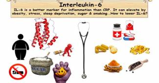
Interleukin-6 (IL-6) is a better marker for inflammation than CRP. This inflammatory cytokine has a strong association with heart disease and asthma.
Interleukin-6 Test
Interleukin-6 is the precursor of C-reactive protein. Thus, IL-6 is a better marker for inflammation than CRP.
Interleukin-6 (IL-6) stimulates the liver to produce a C-reactive protein (CRP). IL-6 is a better marker for inflammation than CRP; because it is the precursor. This inflammatory cytokine has a strong association with heart disease and asthma. Asthma characterized by the airway swelling and constriction; inflammation is behind this.
Interleukin-6 (IL-6) is a cytokine with pro- and anti-inflammatory properties.
After exercise (especially aerobic exercise) IL-6 elevated. If exercise increases IL-6 inflammatory markers; then, how can you say exercise is healthy. Well, when you exercise, your muscles release anti-inflammatory (not inflammatory) IL-6; it is beneficial.
IL-6 levels increased in nearly all sickness and diseases. Your immune cells (macrophages) release pro-inflammatory IL-6. Il-6 is undetectable in the blood or is present in low quantities. Any serum level of ≥ 4.30 pg/mL defined as elevated.
- Optimal: 0 to 14 pg/mL
- High risk: more than 14 pg/mL
Increased levels of IL-6 associated with obesity, type II diabetes, and juvenile rheumatoid arthritis. Different health conditions may elevate IL-6. These conditions are chronic infections, autoimmune disorders, certain cancers and Alzheimer’s disease. Elevated levels of IL-6 associated with an increased risk of heart attack or stroke.
7 Common causes of high IL-6
The most common causes for elevated IL-6 are
- Obesity/overweight,
- Chronic stress,
- Sleep deprivation - too little sleep,
- Eating too much - sugar or refined foods,
- Smoking,
- Excess alcohol drinking,
- Too much of exercise - more than 2 hours of intense exercise per day.
Danger of high IL-6
- IL-6 may cause feelings of “hopelessness”.
- IL-6 may elevate blood sugar levels.
- Studies found IL-6 aggravates the stress hormones (CRH) effects on gut mucosa. This chronic effect leads to Irritable Bowel Syndrome (IBS).
- IL-6 may cause leaky gut.
- People with the major depressive disorder have elevated IL-6. And it raised IL-6, this likely contributes to a worse mood.
- IL-6 is the most potent inducer of C-reactive protein (CRP), an inflammatory marker.
- It creates and worsens food sensitivities and autoimmune issues.
Fasting is not a needed for this test. Can take all prescribed medications, but do not take supplements on the morning of the test.
How do you lower Interleukin-6?
Coenzyme Q10 (CoQ10), omega-3, turmeric, and black pepper help lower your Interleukin-6.
In a trial published on February 16, 2012, in the journal Nutrition. The researchers compared the effects of CoQ10 supplementation. The dosage is 60 or 150 mg/day for 12 weeks; given to men and women with coronary artery disease. "150 mg CoQ10 supplementation appears to decrease the IL-6 in coronary artery disease patients."
Studies show Omega-3 fatty acids inversely related to IL-6. It is useful to lower inflammation and reduce your heart attack risk.
Multi-Ethnic Study of Atherosclerosis found omega-3 intake as un-fried fish was inversely related to IL-6. A study in Norway gives salmon (omega-3 fatty acids) to heart disease patients. The results showed people who ate the high omega-3 containing salmon had a reduction in IL-6. “Omega-3 fatty acids may be both protective; stop inflammation as well as reduce inflammation.”
Omega-3 fatty acids rich foods are
- Oily fish (salmon, herring, mackerel, sardines and tuna),
- Flaxseed, and
- Walnuts.
Antioxidant & anti-inflammatory combination of turmeric & black pepper help lower inflammation. And thus, it lowers IL-6.
"Antioxidant and anti-inflammatory effects of curcuminoid-piperine combination in subjects with metabolic syndrome. A randomized controlled trial and an updated meta-analysis." Published in Clinical Nutrition, December 2015Volume 34, Issue 6, Pages 1101–1108. Short-term curcuminoid-piperine supplementation significantly betterment oxidative and inflammatory status in patients with metabolic syndrome. Curcuminoids could regard as natural, safe and effective CRP-lowering agents.
9 Simple Lifestyle changes to lower IL-6
IL-6 is an inflammatory marker. You can lower IL-6 by avoiding inflammatory and including antioxidant & anti-inflammatory.
The lifestyle modification may have an anti-inflammation effect:
- Reduce or stop eating sugar and refined carbohydrates.
- Eat more antioxidant foods such as berries, cherries, apples, artichokes, dark green leafy vegetables, broccoli, nuts, beans, whole grains and dark chocolates.
- Manage stress with the help of mind-body practices likes yoga, meditation, and Tai Chi. According to a 2010 study, people who do yoga have dropped in interleukin-6 (41%) compared to others. Meditation boosts feel good alpha brain wave activity. Listening relaxation music might be helpful.
- Avoid smoking
- Limit alcohol intake
- Regular moderate exercise.
- Getting optimal sound sleep
- Eating anti-inflammatory foods; pumpkin, sunflower or sesame, nuts such as macadamia and walnuts.
- Plan to take herbs turmeric, ginger, basil, oregano, sage, and thyme.
IL-6 appears to play a role in depression. It suggests that the state of mind plays a role in inflammatory conditions. On this basis, herbs with an antidepressant action may help; you may take St John's wort. Additionally, take anti-inflammatory foods and supplements.
Homocysteine Test
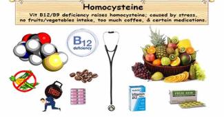
Homocysteine test determines vitamin B9 or B12 deficiency. Vitamin B9 & B12 deficiency linked to increased risk towards a heart attack and stroke.
What is Homocysteine test?
Vitamin B6, B9 or B12 deficiency can cause a rise in homocysteine; risk towards heart attack & stroke. You can normalize it by eating fruit & vegetable or vitamin supplement.
In 1969, Dr. Kilmer S. McCully reported children born with homocystinuria died at a young age due to atherosclerosis. Homocystinuria is a genetical disorder has high levels of homocysteine.
Mildly elevated homocysteine levels are common and seen in 5% to 12% of the general population. Hyperhomocysteinemia is common among alcoholics and patients with chronic kidney disease. High homocysteine associated with low levels of vitamin B6, B12, folate and renal disease.
Homocysteine test determines folate (B9) or B12 deficient. This test diagnoses a rare inherited disorder called homocystinuria. Vitamin B9 & B12 deficiency linked to increased risk towards a heart attack and stroke.
Clinical testing laboratories recommended a healthy homocysteine level is between 5 to 15 µmol/L. But, the Life Extension Foundation believes an upper limit of 15 µmol/L is too high for optimal health. Life Extension recommends a target of lower than 8 µmol/L.
- Optimal: less than 8 µmol/L
- Moderate risk: between 8 to 15 µmol/L
- High risk: more than 15 µmol/L
Danger of high homocysteine
Homocysteine is an amino acid by-product. It causes sticky platelets in blood vessels. Excess homocysteine adversely affects the endothelial function and cardiovascular health.
It also promotes inflammation, oxidative damage, and thrombosis (evil of heart disease). High homocysteine levels can damage the arterial lining. Also, it may increase the chances of blood clot called thrombus.
Thrombus increases the risk of blood vessel blockages. Thrombosis leads to pulmonary embolism, heart attack, and stroke. High levels of homocysteine increase the risk for coronary artery disease.
High homocysteine causes
High homocysteine is highest in people who eat a diet low in fresh fruits, and vegetables. Over or fast cooking destroys the vitamins food contains. Alcohol interferes with folic acid absorption, also increases folate excretion through the urine.
Elevated homocysteine levels may be due to hypothyroidism, kidney disease, psoriasis and some medications. Other contributors to elevated homocysteine are stress and too much coffee consumption. The more coffee you drink, the higher your homocysteine levels are likely to be. The liver metabolizes the stress-induced neurotransmitters epinephrine and norepinephrine. This metabolisis increases the need for folic acid.
High homocysteine is common among people with family history of
- Early heart disease,
- Hypothyroidism, lupus,
- Kidney disease.
Certain drugs tend to elevate homocysteine; they are
- Theophylline (for asthma),
- Methotrexate (for cancer or arthritis),
- L-dopa (for Parkinson’s)
How do you lower Homocysteine?
Take optimal amounts of vitamins B6, B12, B9, and Trimethylglycine (Betaine) to reduce homocysteine.
Luckily, an easy way to bring down the homocysteine level is with three main nutrients Vitamin B6, 9 & 12. Recommended dosages are B9 (folic acid) 400 to 800 mcg, B12 400 to 1,000 mcg, and B6 5 to 20 mg.
“Foliate, homocysteine, endothelial function, and cardiovascular disease. What is the link?” published in Biomed Pharmacother. 2001; 55:425-33. The study proposed to take high doses of folic acid. This supplementation may have beneficial effects on endothelial function also lower homocysteine.
Folic acid supplements and to a lesser extent vitamins B6 & 12 can lower homocysteine levels. In a large population study involving women supplementing high folic acid (as a multivitamin) had fewer heart attacks. Many studies confirm oral folate intake has associated to lower risk of heart disease. This low risk may be due to lowering of homocysteine levels. Eating greener leafy vegetables, orange juice, and beans increases your B vitamins intake.
Animal sources rich in B vitamins are
- Beef,
- Beef Liver,
- Wild Salmon,
- Oats,
- Tuna,
- Turkey Breast,
- Eggs,
- Milk.
Plant sources rich in B vitamins are
- Bananas,
- Potatoes,
- Avocado,
- Kidney Beans,
- Summer Squash,
- Spinach,
- Almonds.
Lifestyle changes to lower Homocysteine
A man too busy to take care of his health is like a mechanic too busy to take care of his tools. ~ Spanish Proverb
- Those who smoked heavily had a 0.8-μmol/liter higher plasma total homocysteine than a nonsmoker. Homocysteine concentrations are 20% higher in smokers than in nonsmokers.
- High levels of alcohol consumption increase plasma total homocysteine concentration. An inverse relation between moderate alcohol consumption and total homocysteine levels has observed.
- Drinking >6 cups of coffee/day linked with a 1.1 to 1.4-μmol/liter higher plasma homocysteine. Drinking 6 cups of strong filtered coffee per day increases homocysteine concentrations. This coffee effect appeared to be due to caffeine and chlorogenic acid it contains.
- A preliminary study found a link between high homocysteine and hostility and repressed anger. The liver metabolizes stress-induced neurotransmitters epinephrine and norepinephrine via a process uses methyl groups. This process increases the need for folic acid. Reduce stress by practicing breathing exercise, meditation, Tai-chi, yoga, music, and bubble bath.
- Increase your B vitamins intake by eating greener leafy vegetables and fruits. The food rich in folate (as folic acid) are green vegetables, orange juice, and beans.
- A study of men with heart disease shown those consuming whole grains and legume at breakfast resulted in a drop in homocysteine. These men take whole grains instead of regular refined rice breakfast.
- An overweight and obese individual have raised plasma homocysteine. Also, reduced vitamin B12 and folic acid levels compared to normal weight individuals.
- High homocysteine may be due to low thyroid hormone, kidney disease, psoriasis and some medications.
Emerging research suggests elevated homocysteine level may be a risk factor for cancer.
Fibrinogen Test
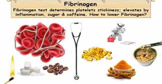
Fibrinogen is a protein in the blood; it determines the platelets stickiness. Fibrinogen is a predictor of cardiovascular risk.
Fibrinogen determines platelets stickiness
Adequate level of fibrinogen is must to stop bleeding when injured. But, an excess of it leads to unnecessary clotting and disturbance of blood circulation.
Fibrinogen is a protein in the blood; it determines the platelets stickiness. During injury, you need adequate fibrinogen level to stop bleeding by forming a blood clot. But, fibrinogen should be within the limit. Otherwise, unnecessary clotting and disturb optimal blood circulation. This test is not preferred; because there is no treatment to lower it if elevated. The normal healthy fibrinogen range is 200 to 400 mg/dL.
Fibrinogen may elevate during inflammation. During the initial phase of the periodontal disease, especially noticeable in gingival tissue. Neonates healthy fibrinogen range is within 125-300 mg/dL (1.25-3 g/L). Fibrinogen levels increase during pregnancy to an average of 4.5 g/l. Fibrinogen level of non-pregnant women is in an average of 3 g/l.
- Optimal: between 150 to 400 mg/dL (1.5 to 4.0 g/L)
- High risk: over 400 mg/dL (>4.0 g/L)
"Dietary Factors Related to Higher Plasma Fibrinogen Levels of Japanese-Americans in Hawaii Compared with Japanese in Japan" published in Arteriosclerosis Thrombosis Vascular Biology. 2006; 26: 1674-1679. Higher intake of iron, sugar, and caffeine along with obesity raises fibrinogen levels.
High Fibrinogen danger
High fibrinogen may increase cardiovascular risk by platelet aggregation, fibrin formation, and plasma viscosity. Elevated fibrinogen level increases the risk of blood clot. Over time, leads to increased risk towards cardiovascular disease.
High Fibrinogen causes
Fibrinogen is must to stop bleeding when injured. Likewise, if there is an internal injury fibrinogen in blood raises to stop in the event of bleeding.
Elevated levels of Fibrinogen may be due to
- Acute infection,
- Cancer,
- Coronary heart disease & myocardial infarction
- Stroke,
- Inflammatory disorders (rheumatoid arthritis and glomerulonephritis - a form of kidney disease),
- Trauma,
- Cigarette smoking,
- Pregnancy, and
- Peripheral artery disease.
Fibrinogen elevation is common among cigarette smokers. It has been identifying as a primary mechanism causing heart disease and stroke. Cigarette smoking increases cardiovascular disease risk and raises fibrinogen levels. Quitting smoking might help slight reductions of fibrinogen levels.
How can you lower Fibrinogen? 9 nutrients are helpful!
Nattokinase acts like blood thinner; thus, it prevents unwanted clotting and lower fibrinogen.
“Nattokinase decreases plasma levels of fibrinogen, factor VII, and factor VIII in human subjects” published in Nutrition Research 2009 Mar;29(3):190-6. doi: 10.1016/j.nutres.2009.01.009. Natto is a traditional Japanese food made from soybeans fermented with Bacillus subtilis. Nattokinase is a serine protease from Bacillus subtilis. It is considering as one of the most active functional ingredients found in natto. Nattokinase could reduce certain factors of blood clotting and lipids. In summary, this study showed oral nattokinase could lower plasma fibrinogen level and CVD.
Studies show Omega-3 fatty acids has hypo-coagulant property. It is useful to lower fibrinogen and reduce your heart attack risk.
Omega-3 fatty acids may also help when there is high fibrinogen. Taking 3000 mg/day of fish oil shown to lower fibrinogen levels. Fish oil shows to reduce heart attack risk. Additionally, reduce triglycerides, suppress inflammation, decreased platelet stickiness, and protect against arrhythmia.
"Variable hypo-coagulant effect of fish oil intake in humans: modulation of fibrinogen level and thrombin generation" published in Arteriosclerosis Thrombosis Vascular Biology. 2004 Sep;24(9):1734-40. Epub 2004 Jun 24. Intake of fish oil reduced fibrinogen, factor V levels, and thrombin generation in plasma. Thus, dietary fish oil can provoke a hypo-coagulant effect depending on the fibrinogen level.
Differential effects of dietary supplementation with fish oil or soy lecithin on human platelet adhesion. Thrombosis Haemostasis 1999 Nov;82(5):1522-7.
Studies have demonstrated Niacin (vitamin B3) can help lower fibrinogen.
If you can tolerate unpleasant side effects of niacin, then niacin can help lower fibrinogen. "Effect of niacin supplementation on fibrinogen levels in patients with the peripheral vascular disease." American Journal of Cardiology 1998 Sep 1;82(5):697-9, A9. "Nicotinic acid treatment shifts the fibrinolytic balance favorably and decreases plasma fibrinogen in hyper-triglyceride men." Journal of Cardiovascular Risk 1997 Jun;4(3):165-71.
Vitamin C lower your coronary artery disease risk by decreasing platelet aggregation.
Vitamin C supplementation of 2000 mg/day can increased fibrinogen breakdown by 45%. Additionally, it reduces platelet aggregation by 27% and total cholesterol by 12%. Ref: "The effect of vitamin C on blood lipids, fibrinolytic activity and platelet adhesiveness in patients with coronary artery disease." Atherosclerosis. 1980 Feb;35(2):181-7.
Anti-inflammatory Turmeric can lower high fibrinogen.
Eight subjects with elevated fibrinogen levels treated with 20 mg/day of turmeric extract. After 15 days, fibrinogen dropped in all eight subjects. "A hydroalcoholic extract of Curcuma longa lowers the abnormally high values of human plasma fibrinogen" published in Mechanisms of Ageing and Development 2000, 114: 207-220.
Green tea, ginkgo, garlic and vitamin E have an inhibitory effect on platelet aggregation.
References:
- "Antithrombotic activities of green tea catechins and (-)-epigallocatechin gallate." Published in Thrombosis Research 1999 Nov 1;96(3):229-37.
- "Actions of Ginkgo Biloba related to potential utility for the treatment of conditions involving cerebral hypoxia." Published in Life Sciences 2000 Aug 11;67(12):1389-96.
- "Dietary supplementation with aged garlic extract inhibits ADP-induced platelet aggregation in humans." Published in Journal of Nutrition 2000 Nov;130(11):2662-5.
- "Specific cellular responses to alpha-tocopherol." Published in Journal of Nutrition 2000 Jul;130(7):1649-52.
Maintaining blood sugar A1C below 4.6% can lower your risk towards coronary heart disease.
Contrary to conventional wisdom, we advised trying to keep their glucose levels at 85 mg/dL or lower. Based on recent findings, an ideal reading of hemoglobin A1C is 4.5% or lower. Ref: "Glycemic control and coronary heart disease risk in persons with and without diabetes: the atherosclerosis risk in communities’ study". Published in Archives of Internal Medicine 2005 Sep 12;165(16):1910-6.
The proteolytic enzyme bromelain may be the most effective fibrinogen-lowering supplement.
CRP Test
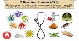
C-Reactive Protein is a marker for inflammation. CRP has identified as a reliable predictor of heart health than elevated cholesterol levels.
C-Reactive Protein Test
In many studies, CRP identified as a reliable heart health predictor than cholesterol levels.
This protein is a marker for inflammation associated with cardiovascular health. High CRP level is an indicator of infections, high sugar, excess weight, and hypercoagulability. Make sure to use the high-sensitivity test (hs-CRP). The recommended optimal CRP level is lower than 0.8 mg/dL.
High-sensitive CRP
- Optimal: less than 1 mg/dL (<10 mg/L)
- Moderate risk: between 1 to 3 mg/dL (10 to 30 mg/L)
- High risk: more than 3 mg/dL (>30 mg/L)
Inflammatory processes are independent predictors of heart health risk. Test reveals the level of blood vessel inflammation. In due course, leads to plaque rupture that result in heart attack or stroke.
CRP causes
Increased C-reactive protein (CRP) level has an associated with:
- Cardiac risk factors,
- Advanced age,
- Diabetes mellitus,
- Elevated cholesterol,
- Increased body mass index (BMI) & obesity,
- Cigarette smoking.
High C-reactive protein (CRP) levels might be due to:
- Infection,
- Inflammatory bowel disease (IBS),
- Lupus,
- Pneumonia,
- Rheumatoid arthritis,
- Tuberculosis,
- Heart attack,
- Connective tissue disease,
- Cancer.
How can you lower CRP?
Studies suggest, you can lower CRP with simple lifestyle change and taking herbal supplements.
To lower CRP, various studies recommend to:
- Limit high glycemic foods,
- Avoid gluten,
- Take antioxidant such as turmeric & Amla
- Consume anti-inflammatory black pepper & ginger.
Ref: “Relation between a Diet with a High Glycemic Load and Plasma Concentrations of High-Sensitivity CRP in Middle-Aged Women.” Published in American Journal of Clinical Nutrition 75, no. 3 (2002): 492–98. Researchers assessed 244 healthy women diets to check association between glycemic load and CRP. High glycemic diet had twice the CRP; compared with lowest glycemic. Overweight women had high inflammation levels.
Whole grains raise blood sugar as much as processed grains, may be at a slow pace. Gluten it contains; causes inflammation to gluten sensitive people. Avoid or at least reduce, consumption of processed carbohydrates. Instead, increase unprocessed carbohydrates from vegetables and low sugar fruits.
Antioxidant and anti-inflammatory combination of turmeric & black pepper help lower inflammation thus CRP.
"Antioxidant and anti-inflammatory effects of curcuminoid-piperine combination in subjects with metabolic syndrome. A randomized controlled trial and an updated meta-analysis." Published in Clinical Nutrition, December 2015Volume 34, Issue 6, Pages 1101–1108. Short term curcuminoid-piperine supplementation betterment oxidative and inflammatory status in patients with metabolic syndrome. Curcuminoids could be a natural, safe and effective CRP-lowering agents.
Studies shows Amla is a strong antioxidant. It is useful for your heart and blood vessels health.
Studies consistently shows amla extract improves HDL. Reliable studies show amla additionally lowers triglycerides as well as C-Reactive Protein (CRP). Amla has incredible antioxidant properties, useful for your heart and blood vessels. Amla limits the cholesterol oxidation in your arteries. It is crucial for a long and healthy life.
Ginger contains potent anti-inflammatory compounds called gingerols.
In February 2005, A study published in the Journal of Alternative and Complementary Medicine. This study sheds light on the mechanisms of action that underlie ginger's anti-inflammatory effectiveness. In this research, ginger shown to suppress pro-inflammatory compounds cytokines and chemokines. These pro-inflammatory is from cell lining of the joints, joint cartilage and immune cells.
"Anti-Inflammatory Effects of Zingiber Officinale in Type 2 Diabetic Patients." Published in Advanced Pharmaceutical Bulletin 2013 Dec; 3(2): 273–276. Ginger supplementation significantly reduced the levels of TNF-α, IL-6, and hs-CRP.
"Ginger and its Effects on Inflammatory Diseases." Published in Advances in Food Technology and Nutrition Sciences Open Journal 2015; 1(4): 97-101. Ginger can help in the treatment of inflammatory-chronic-diseases. Such inflammatory conditions are Fatty Liver, Asthma, Cancer and Arthritis. Ginger help through anti-inflammatory, immune regulatory and anti-oxidative mechanisms.
8 Simple Proved Lifestyle changes to lower CRP
CRP is the inflammatory marker. To lower CRP, you have to avoid inflammatory, and add antioxidant & anti-inflammatory.
- If you have any inflammatory health condition, then proper treatment is a must. Some inflammatory conditions are Allergy, Asthma, Arthritis, Celiac, Crohn's Disease, Eczema, Fibromyalgia, Fibrosis, GERD, Lupus, Neuropathy, Pancreatitis, Psoriasis, Rheumatoid Arthritis, etc.
- Overweight and obese people tends to have higher levels of CRP. Bigger fat cells excrete more interleukin-6 (IL-6). This IL-6 triggers the liver to produce more CRP.
- Smoking hardens arteries as well as raise CRP levels. Research shows, you can reverse arterial damage within ten years of quit smoking.
- Anti-inflammatory foods are alpha-linolenic acid rich flaxseed, walnuts & canola oil. Additionally, omega-3 fats in certain fish fight inflammation. Eat food more of fresh fruits, vegetables, whole grains, nuts, and olive oil. It can lower inflammation and decrease CRP levels. Eating more fish or taking fish oil supplements can reduce C-reactive protein levels. Consuming dark chocolate can reduce CRP levels. The highest fiber intake lower CRP. Spices such as Turmeric, Ginger & Garlic are helpful. Herbal teas especially green tea can be useful to reduce inflammation.
- Avoid inflammatory foods such as sugar, red meat, refined flours, dairy, and trans fats. Limit or stop products made out of refined flours such as white bread and pasta. You should avoid dairy, although yogurt and kefir are good for your guts. Soft drink, caffeine, and alcohol can product inflammation, thus avoid it.
- Get enough sleep, because sleep deprivation increase risk for stroke or heart attack. People with sleep deprivation can even suffering from sudden cardiac death.
- High levels of stress hormones lead to the release of excess inflammatory chemicals. Reduce stress by practicing 15 minutes of relaxation. Best relaxations are deep breathing, meditation, yoga, Tai Chi, music, or a bubble bath.
- Exercise is essential to help lower cortisol levels. Thus, reduce inflammation in the body. But, choose the right exercise for your body is important. Over doing exercise may elevate inflammation.
Reflexology Skeletal System
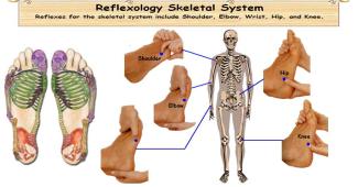
Human skeleton consists of three sections; bones, cartilages, and joints. Bone is a hard pattern of connective tissue. Cartilage is also a connective tissue; however, not as hard as a bone. Joints in the human skeleton enable movement. Skeletal system comprises 206 bones; vertebral column, breastbone, shoulder blades, collarbones, elbow, wrist, metacarpals, phalanges, pelvic girdle, thigh, knee, shin, and foot bones.
5 Sturdy reflexes Strong Skeletal
The job of the human skeleton is to provide strength, support, and shape. In addition, it protect delicate organs and gives mobility.
Teeth is part of the skeletal system made of enamel and dentin; however, not consider as a bone. Enamel is the strongest substance in the body.
Spinal cord consists of 26 vertebrae, forming the human vertebral column. Neck contain seven cervical vertebrae, Chest contains 12 Thoracic vertebrae, Lower back contain five Lumbar vertebrae, Sacrum contains one vertebra, and Coccyx or tailbone contain one vertebra.
Shoulder reflex point
How to do reflexology therapy for the shoulder? On the fourth and fifth toe base, place the index finger on the dorsal aspect and the thumb on the lateral aspect. Walk four steps away from the toe. Reflexology therapy for the shoulder is useful for shoulder problems; it relieves tension as well as facilitates mobility.
Wrist reflex point
How to do reflexology therapy for the wrist? Using the thumb, walk from the fifth toe base on the lateral aspect one-step lower. Apply pressure for 5 seconds. Reflexology therapy for wrist is useful for carpal tunnel syndrome and wrist injury.
Elbow reflex point
How to do reflexology therapy for the elbow? Using the thumb, walk from wrist reflex one-step towards the elbow reflexes. Apply pressure for 5 seconds. Reflexology therapy for elbow is useful for nerve problems in the arms, elbow arthritis, tennis elbow, and faster fracture healing.
Knee reflex point
How to do reflexology therapy for the knee? Using the thumb, walk two steps from elbow reflexes below to reach a small bony protrusion on the foot lateral side. Hook and backup for 5 seconds. Reflexology therapy for the knee is useful to reduce knee pain, ligament sprain, arthritis, and strengthen knee joint.
Hip reflex point
How to do reflexology therapy for the hip? Using the thumb, walk diagonally two steps towards anklebone from the knee reflexes. Apply pressure for 5 seconds. Hip reflexology therapy is useful for arthritis, heal fractures, hip pain, and faster recovery from hip surgery.
Reflexology Reproductive System

The reproductive system contains a set of sex organs; it works together for the sexual reproduction. The male reproductive cells important job is to fertilize female reproductive cell egg.
4 Cheerful Reflexes for Foolproof Reproduction
- The male reproductive system comprises the penis, the testicles, the epididymis, the vas deferens and the prostate gland. Androgens are hormones make men ‘male’. The sperm is the male reproductive cell, the important job is to fertilize an egg.
- The female reproductive system consists of the vagina, womb (uterus), Fallopian tubes and ovaries. A female’s entire egg develops, even when she is an unborn baby. During puberty, the eggs ripen inside the ovary and release every month.
Uterus (in female) Prostate (in male) reflex point
Uterus known as Womb is a hollow muscular organ of the female reproductive system; it develops the embryo and fetus during pregnancy.
A walnut-sized gland prostate secretes sperm; it is a protecting and nourishing fluid. Prostate located in front of the rectum between the bladder and the penis; additionally, the urethra extending from its center.
How to do reflexology therapy for the Uterus or Prostate? Uterus or Prostate reflexes are on the medial side, halfway between the back of the heel and the anklebone. Using the index finger, press and rotate this point for ten seconds. Uterus and prostate reflexology therapy are helpful for good general reproductive health.
Ovary (in female) Testicle (in male) reflex point
Reproductive gland of the female is ovaries located in the pelvis. It secretes hormones estrogen and progesterone; it is responsible for puberty, menstrual cycle control, and pregnancy.
Reproductive gland of the male is testes located in the scrotum. It secretes hormone testosterone. Testosterone is responsible for puberty; manages height growth, deepening voice, facial & pubic hair growth, and sperm production.
How to do reflexology therapy for the Ovary or Testicle? Ovary or Testicle reflexes are on the lateral side, halfway between the back of the heel and the anklebone. Using the index finger, press and rotate this point for ten seconds. Ovary or testicle reflexology therapy help produce sex hormones, regulate ovulation, and healthy sperm production.
Fallopian tube (in female) Vas deferens (in male) reflex
In the female, the fallopian or uterine tube delivers the ova from the ovary to the uterus every month. Sperm fertilizes ovum; after fertilization, the uterine tube carries fertilized egg to the uterus.
In the male, the vas deferens is a long muscular tube carry mature sperm to the urethra for ejaculation.
How to do reflexology therapy for the fallopian tube or Vas deferens? Using the index and middle finger, walk on the dorsal (top) aspect; from lateral ankle bone to medial ankle bone and walk back. Repeat three times. Reflexology therapy for the fallopian tube or Vas deferens is useful for fertility related issues.
Breast (in female) reflex
The breast is medically referred as the mammary gland; it is a milk-producing structure composed of fat cells.
How to do reflexology therapy for the Breast? Using three fingers on the dorsal aspect (topside) of the foot; halfway up the foot between zone 4 & 5 to make the large rotary motion. Repeat five times. Reflexology therapy for breast is helpful for breast disorders including infection, tumors, and tenderness.
Alternative method: Using three finger on the dorsal aspect of the foot; walk from lateral to medial between zone 5 to 3.
For disorders of reproduction also, add pituitary and adrenal glands for extra benefit.
Reflexology Excretory System
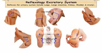
The excretory system removes excess, unnecessary materials from the body; so helps to keep the internal chemical homeostasis and prevent damage to the body.
5 Unique Organ Reflexes for proper Excretion
Reflexes for the urinary system include kidney, ureters, bladder, and urethra.
- The urinary system comprises kidney, ureters, bladder, and urethra. The kidneys essential jobs are to remove waste products (urea, salts, and excess water) from the blood and regulate the fluid balance. Ureters are muscular ducts; from the kidneys, it propel urine to the urinary bladder. Urinary bladder collects waste (urine) from the kidneys. The urethra is a tube connects the urinary bladder to the external body.
- Throughout the body, skin contains sweat glands; it secretes sweat. Reflexology therapy for digestive system additionally nourishes skin and sweat glands.
- The lungs expel gaseous wastes (e.g. carbon dioxide). Lungs reflexology therapy is in the respiratory system.
Kidney and Adrenal reflex points
Two bean-shaped organs called kidney located on either side of the spine. Important jobs of the kidneys are to remove waste from the blood (urea, salt, and excess water), maintain body's fluid balance, control blood pressure, govern red blood cells, and acid neutralization.
The kidney reflexes are on right and left feet; located at the waistline near the base of the second metatarsals. Similar to the kidney's location in the body, the kidney reflexes are on either side of the spine reflexes; right side kidney below liver is lower than the left side.
How to do reflexology therapy for the kidney? Using two thumbs together, pull apart with a gentle pressure to reach the kidney and adrenal reflexes. Do for 10 seconds.
Rotate the kidney reflex point with the right thumb. Work on this reflex helps cut body pain and support body to cope with stress.
Using the right thumb, rotate the kidney reflex point. Reflexology therapy for kidney helps to relieve body pains and cope with stress.
Ureter tube reflexes
Ureter consists of two 10 to 12 inches long uterine tubes; from the kidneys, it moves the urine to the bladder, using gravity and waves of contractions.
How to do reflexology therapy for the ureter? Using the left thumb, walk downwards from the kidney reflexes until reaching the bladder reflexes. Repeat three times. Reflexology therapy for ureter is useful for bed-wetting, local inflammation, water retention, and kidney stones.
Bladder reflex
The urinary bladder is a pear-shaped muscular sac; located above and behind the pubic bone. The bladder stores urine as well as allows occasional and voluntary urination.
How to do reflexology therapy for the bladder? Using the thumb; walk from the bladder reflex spread outwards (30 degrees apart gap), like bicycle spokes. Repeat five times. Patients with cystitis contain crystals in this reflex. Apply pressure on the bladder reflex; help to disperse these crystals. Need to drink more water for safe flush out of these crystals.
Urethra reflex point
The urethra is a tube to expel urine from the urinary bladder. In males, the urethra works as a passage for cells and secretions from various reproductive organs.
How to do reflexology therapy for the urethra? Using the thumb, walk from bladder reflexes until reaching the heel bone center. Repeat three times.