
Dry Eye
Dry eye syndrome is an eye problem with low quantity or quality of tears that make it dry and red. Detail info of its causes, symptoms, test, and treatments.
Dry eye is a common and often chronic problem, particularly in older adults. It is a condition in which there are insufficient tears to lubricate and nourish the eye. Need tears for maintaining the health of the front surface of the eye and for providing clear vision?
With each blink of the eyelids, tears are spreading across the front surface of the eye, known as the cornea. Tears provide lubrication, reduce the risk of eye infection, wash away foreign matter in the eye, and keep the surface of the eyes smooth and clear. Excess tears in the eyes flow into small drainage ducts, in the inner corners of the eyelids, which drain in the back of the nose.
Dry eye syndrome
Dry eyes can result from an improper balance of tear production and drainage.
Inadequate amount of tears - several glands in and around the eyelids produce Tears. Tear production tends to diminish with age, with various medical conditions, or as a side effect of certain medicines. Environmental conditions such as wind and dry climates can also affect to tear volume by increasing tear evaporation. When the normal amount of tear production decreases or tears evaporate too quickly from the eyes, symptoms of the dry eye can develop.
Poor quality of tears - Tears are of three layers: oil, water, and mucus. Each component serves a function in protecting and nourishing the front surface of the eye. A smooth oil layer helps to prevent evaporation of the water layer, while the mucus layer helps in spreading the tears evenly over the surface of the eye. If the tears evaporate too quickly or do not spread on the cornea due to deficiencies with any of the three tear layers, dry eye symptoms can occur.
The most common form of dry eyes is due to an inadequate amount of the water layer of tears. This condition, called kerato conjunctivitis sicca (KCS), is also referring to as dry eye syndrome.
People with this condition may experience eye irritated, and advanced stage may damage the front surface of the eye and impair vision. Proper treatments for dry eyes aim to restore or maintain the normal amount of tears in the eye to minimize dryness and related discomfort and to maintain eye health.
Strabismus Treatments
People with strabismus have several treatment options available to improve eye alignment and coordination. They include:
Strabismus treatment options
- Eyeglasses or contact lenses – suitable for accommodative esotropia
- Prism lenses
- Vision therapy
- Eye muscle surgery
Eyeglasses or Contacts lenses
Eyeglasses or contact lenses may prescribe for patients with uncorrected farsightedness. This may be the only treatment needed for some patients with accommodative esotropia. Once the farsightedness is correct, the eyes require less focusing effort and may remain straight.
Prism lenses
Prism lenses are special lenses that have a prescription for prism power in them. The prisms alter the light entering the eye and assist in reducing the amount of turning the eye has to do to look at objects. Sometimes the prisms are able to compensate for and eliminate the eye turning.
Vision therapy
Vision therapy is a structured eye-training program of visual activities suggested to improve eye coordination and eye focusing abilities. Vision therapy helps and trains the eyes and brain to work together more effectively. These eye exercises help remediate deficiencies in eye movement, eye focusing, eye teaming, and reinforce the eye brain coordination or connection. Treatment may include office based as well as home-based training procedures.
Strabismus surgery – eye muscle surgery
Eye muscle surgery can change the length or position of the muscles around the eye in an attempt to align the eyes. Eye muscle surgery is performing to align the eyes, so they appear straight. After surgery, a vision therapy is often requiring for effective eye coordination and to keep the eyes from revert to their previous condition of misalignment.
Strabismus Diagnose
Strabismus is diagnosing through a comprehensive eye exam. Testing for strabismus, with special emphasis on how the eyes focus and move, may include:
Strabismus diagnosis test
- Patient History – Need a patient history to determine any symptoms the patient is experiencing, presence of any general health problems, medications taken, or environmental factors that may be contributing to the symptoms.
- Visual Acuity - Visual acuity measurements are taking to assess the extent to which the vision may be affected. As part of the testing, patients will ask to read letters on specific distance and near reading charts. This test measures visual acuity, which is writing as a fraction such as 20/40. When testing distance vision, the top number is the standard distance at which testing is performing, twenty feet. The bottom number is the smallest letter-size that can read at the twenty-foot distance. A person with 20/40 visual acuity would have to get within 20 feet of a letter that should see at forty feet in order to see it clearly. Normal distance visual acuity is 20/20.
- Refraction - it is conduct to determine the appropriate lens power needed to compensate for any refractive error (nearsightedness, farsightedness, or astigmatism). Using an instrument called a phoropter, the optometrist places a series of lenses in front of the eyes, and measures how they focus light using a hand held lighted instrument called a retinoscope. Alternatively, the doctor may choose to use an automated instrument that automatically evaluates the refractive power of the eye. The power is then refining by the patient's responses to determine the lenses that allow the clearest vision.
- Alignment and Focusing Testing - How well the eyes focus, move and work together needs to assess. In order to obtain a clear, single image of what is being viewing, the eyes must effectively change focus, move, and work in unison. This testing will look for problems that keep the eyes from focusing effectively or make it difficult to use both eyes together.
- Examination of eye health - The structure of the eye is observing to rule out any eye disease that may be contributing to strabismus. The health of the external and internal parts of the eye will assess using various testing procedures.
This testing may be without the use of eye drops to determine how the eyes respond under normal seeing conditions. In some cases, such as for patients who cannot respond verbally or when some of the eyes focusing power may hide; eye drops may be used. They temporarily keep the eyes from changing focus while doing this test.
Using the information obtained from these tests, along with results of other tests, the optometrist can determine if one have strabismus. Once testing is complete, the optometrist can discuss options for treatment.
Strabismus Symptoms
Strabismus or crossed eye causes may be due to muscular, nerve or other general health problems discuss in detail.
What causes strabismus?
- Eye muscle problems,
- Nerve problem that transmits information to the muscles,
- Problem with the brain's control center that controls the eye movements
- Other general health conditions or eye injuries
Risk factors for developing strabismus
- Family history - individuals with parents or siblings who have strabismus are more likely to develop it.
- Refractive error - people who have a significant amount of uncorrected farsightedness (hyperopia) may develop strabismus, because of the additional amount of eye focusing required to keep objects clear.
- Medical conditions - people with conditions such as Down syndrome and cerebral palsy. Alternatively, who have suffered with a stroke or head injury.
Although there are many types of strabismus that can develop in children or adults, the two most common forms are accommodative esotropia and intermittent exotropia.
Esotropia and its symptoms
Accommodative esotropia mostly occurs because of uncorrected farsightedness or hyperopia. Because the eyes focusing system is linked to the system that controls where the eyes point, the extra focusing effort needed to keep images clear in farsightedness may cause the eyes to turn inward.
Signs and symptoms of accommodative esotropia may include seeing double vision, urge to close or cover one eye when doing close work, and tilting or turning of the head.
Exotropia and its symptoms
Intermittent exotropia may develop due to an inability to coordinate both eyes together. The eyes may have a tendency to point beyond the object being viewing.
Signs and symptoms of intermittent exotropia may include headaches, difficulty reading, and eyestrain. They also may have a tendency to close one eye when viewing at distance or in bright sunlight.
Strabismus

Crossed eye or strabismus is mistakenly considering by many as Lazy eye or Amblyopia, but amblyopia and strabismus are different.
The confusion between these two conditions may be because a cross eye can cause lazy eye. In other words, a constant unilateral crossed eye (strabismus) can cause lazy eye (amblyopia). However, infrequent crossed eye (strabismus occurs rarely) may rarely cause lazy eye.
Strabismus crossed eye
Strabismus crossed eyes is a condition of both eyes do not look at the same place at the same time. It occurs when an eye turns in, out, up or down and is usually causing by poor eye muscle control or a high amount of farsightedness.
There are six muscles attached to every eye that control how it moves. The muscles receive signals from the brain that instructs their movements. Normally, the eyes work together so they both point at the same place. When there is any problem develops with eye movement control, then an eye may turn in, out, up, or down. The eye turning may occur more frequently or may appear only at certain conditions such as when the person is tired, ill, or has done a lot of reading or close work. In some cases, the same eye may turn each time, while in other cases, the eyes may alternate turning.
A proper eye alignment is important to avoid seeing double vision, for good depth perception, and to prevent the development of poor vision in the turned eye. When the eyes are misaligned, the brain receives two different images. At first, this may create double vision and confusion, but over time, the brain will learn to ignore the image from the turned eye. If the eye turning becomes constant (frequent) and is not treated properly, it can lead to permanent vision reduction in one eye, a condition called amblyopia or lazy eye.
Some baby’s eyes may appear to misaligns, but are actually both aim at the same object. This is a condition called pseudo-strabismus or false strabismus. The appearance of crossed eyes may be due to extra skin that covers the inner corner of the eyes, or a wide bridge of the nose. Usually, this will change as the child's face begins to grow.
Strabismus usually develops in infants and young children, mostly at the age of three, but older children and adults can also develop the condition. There is a common misconception that a child with strabismus will outgrow the condition, but this is not true. Actually, strabismus may become worse without proper treatment. Any child older than four months whose eyes do not appear to be straight all the time should examine for strabismus.
Strabismus types or classification
- Inward turning known as esotropia
- Outward turning known as exotropia
- Upward turning known as hypertropia
- Downward turning known as hypotropia
Other classifications of strabismus include:
- The frequency with which it occurs - either constant or intermittent
- It always involves the same eye (only one eye) - unilateral
- It involves both eyes (right eye or sometimes left eye) - alternating.
Reflexology After Care
Improvise reflexology health benefit from the reflexology treatment by trying to have rest and take things easy as this helps to boost the body's natural healing abilities.
Possible effects after reflexology treatment
After-effects of reflexology are unique to an individual person; most of them experience a sense of well being, relaxation, uplifting, and energizing.
Few may feel tiredness, nauseous, cold symptoms, urinate more frequently, or more emotional, or too lethargic this called as a healing crisis. It is an indication of the body start eliminating toxins and heals itself.
After Care Advice
Drink more water than normal can help to flush the toxins out of the system thereby aiding the detoxifying process. It is also recommended that to avoid alcohol and eat only light meals (avoid heavy meals immediately after treatment).
After one or two reflexology treatments, the body starts to respond in a very definite way. Most people experience a sense of well-being and relaxation; sometimes people report feeling of lethargy, nausea or tearful, but this is transitory and is a part of the holistic healing process.
Does reflexology cause any side effects?
No, it does not cause any undesirable permanent side effects or make any condition more acute. Reflexology helps to normalize body functions. A Reflexology session relaxes the stress that can affect the health. It is a safe natural way to better health.
Improvise reflexology health benefit from the reflexology treatment by trying to have rest and take things easy as this helps to boost the body's natural healing abilities.
Colorblindness Treatments
Colour blindness treatment can be of many ways just to reduce the symptoms of color blindness. Best thing is to learn to live with color blindness.
Color blindness correction
There are many things that doctors can do to reduce the symptoms of color blindness. One is to use color filters such as a red-orange filter helps to interpret colors or to see contrasts. Red filters can absorb short and medium wavelengths of the light spectrum, leaving only the longer ones.
Another way to reduce color deficit symptoms is to use X-Chrom lenses. The X-chrom lens is a red contact lens, which is normally warning on the non-dominant eye of color deficient people that help to interpret colors or their contrasts. It has proven to help patients with the ishihara test, but has made it either harder for the color deficit patient to take other tests or the test results remain the same as without the lens. Therefore, X-Chrom lenses may help the patient with everyday hobbies but should not consider as having no color deficient.
Tips for people with color blindness
- Label items that require color recognition.
- Differentiate colors based on location (for example, traffic lights)
- Maximize color contrast if possible.
- Label coloring utensils (crayon, pen, pencil) with the color name.
- Organizing and labeling clothing to avoid colors that clash with one another.
- Specially tinted glasses can assist some individuals to tell the difference between certain colors. Although these glasses assist, they do not restore normal color vision.
Reflexology Hand Chart
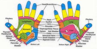
Hand Reflexology is equally effective as foot reflexology. There are reflex points on the hands indicating every organ, gland, and parts of the body.
Hand Reflexology
By using the unique thumb and finger techniques on these hand reflexes, the body responds by relaxing, rebalancing, and improving blood circulation. The hands are not as sensitive as the feet, and the reflex points are much more profound. However, a hand reflexology treatment can be worthwhile.
Hand reflexology can use as a stand-alone treatment in its own right. When the foot has injured or other problems that can stop performing foot reflexology.
Are you lazy doing reflexology? You can do foot tapping. It is easy to do with numerous health benefits; it improves your overall health profile. You can learn more by visiting foot tapping.
You may also interest to know the benefits of reflexology massage, how to do? And reflexology aftercare.
Colorblindness Test
Colour-blind test mostly performed using ishihara plates with pictures of colored spots easily distinguished by normal person, but not by colorblind.
Ishihara Color Test
Most commonly, color blindness is testing by using Ishihara Color Test. Dr Shinobu Ishihara, professor in University of Tokyo, designed series of pictures of colored spots, which can use to diagnose red green color blindness. In the image below, they contain a series of Ishihara pictures. We have prepared a color blindness test using these Ishihara test plates’ tests yourself, or someone you love, might be color blind – or see it just for fun!
A numbers shape or image is embedding in the picture as a number of spots in a slightly different color, which can see with normal color vision. However, cannot by seen and distinguish the number (or shapes or image) embedded in the picture by persons with a particular color defect. The full test set has a variety of figures embedded in the color combinations, and enable diagnosis of which particular visual defect is present.
Ishihara color test contains only numerals; it becomes difficult to diagnose a young child, ho does not yet learn numbers. To identify color blindness early on in life, alternative color vision tests were design using only symbols (square, circle, & car) that are known to children.
Have a free Ishihara color blindness online test
This online color vision test consists of ishihara color plates with an embedded number. Take 3 seconds to identify the number on each of the plates. To know the actual hidden number, just click on the image to see the answer.
.
Ishihara colour test plate 1

Ishihara colour test plate 2

Ishihara colour test plate 3

Ishihara colour test plate 4

Ishihara colour test plate 5

Ishihara colour test plate 6

Ishihara colour test plate 7

Ishihara colour test plate 8

Ishihara colour test plate 9

Ishihara colour test plate 10

Ishihara colour test plate 11

Ishihara colour test plate 12

Ishihara colour test plate 13

Ishihara colour test plate 14

Ishihara colour test plate 15

If able to recognize the hidden numbers in the above 15 ishihara color-blindness plates, then congratulations, you are not colorblind. If not able to recognize some of the hidden numbers, then may be color blind go to doctor’s office to confirm. Read further to know how to lead a convenient life with color blindness.
Colorblindness Causes
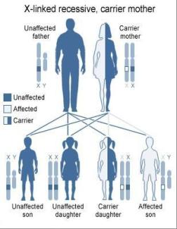
We do not know the causes of color blindness, but most of the causes of color blindness are inheriting from birth, that is genetics play a major role.
Causes of Color deficiency or blindness
- Most of the case’s color blindness is inherited (Possessed at birth).
- Some time it may be due to an injury.
- Some times aging process play a role.
- Other health conditions (diseases) may trigger it.
- Rarely may it cause by the side effects of any medications.
Eye’s retina has cones (light sensitive cells), which are responsible for sensing colors based on its wavelength. There are three different types of cones to sense three colors (red, green, and blue) and their combinations or shades.
Color blindness occurs when cones (or light sensitive cells) in the retina fail to sense properly to the variations in wavelengths of light that enters the eye. These are causes due to the absence of cones in the retina or if these cones not responding to the color of lights.
Other conditions that may cause color blindness:
Parkinson's disease (PD) is a neurological disorder, in which the light-sensitive nerve cells in the retina may damage and cannot function properly.
Genetically caused color blindness
The most common congenital color vision defect is the red green deficiencies, which are due to "X chromosomes (decides baby sex)." Men are mostly affecting because women have two X chromosomes, and men have only one X and a Y chromosome. If a man's one X chromosome is color defective, he will be color deficient, whereas in a woman must have both the X chromosomes color deficient, then only she will be color deficient. If one X chromosome is color defective and the other is not, then she is not a color deficient but a carrier of color deficient. For a woman to be color deficient her father must be colorblind, and her mother is colorblind or carrier of color blind.