
Eye Infection
Eye infection is a pathological state of eye resulting from the invasion of pathogenic microorganisms, such as virus, bacteria, and fungus.
Infections of Eye
The eyes are frequently exposing to various pathogenic microorganisms. When the normal required defense (immune) mechanism of the eye fails to protect, may end up with eye infections. The source of the infection may be local that is from the eyelids or remote that is from the sinuses. Infection can also be the caused due to trauma, eye surgery, contact lens usage, or other diseases resulting in bacteria growth or viruses.
Cleaner Eye - Eye tears and eye lid
Normally eyes are protecting from dust and pathogens by flashing the eye surface with eye tears and gently washed it or wipe it with eye lips. This helps to remove dirt and pathogens that may affect the front surface of the eye and protects from eye infection.
Eye infections can affect any age group, but those who had eye-surgery or experienced an eye trauma are at higher risk for infection. Few eye infections are highly contagious, such as pink eye, those who were exposing to someone who had infected are at a much higher risk to develop infection.
Retinitis Pigmentosa
Retinitis pigmentosa (RP) is the degeneration of the light-sensitive retina that progressively leads to night, peripheral, and then central vision loss.
Retinitis pigmentosa
Retinitis pigmentosa (RP) is an inherited eye disorder characterized by progressive peripheral vision loss, followed by night-vision difficulties and then leads to central vision loss. Vision loss is due to slow degeneration of light sensitive cells (rods & cones) in the retina.
Retinitis pigmentosa symptoms
The first symptom of retinitis pigmentosa usually occurs in early childhood; mostly, both eyes are affecting with poor night vision, followed by narrowing of the field of vision. At its later stages of retinitis pigmentosa, only a tiny area of central vision exists called as tunnel vision, along with some little peripheral vision.
Retinitis pigmentosa causes
Causes of retinitis pigmentosa are unclear, except that inherit. Hereditary is blame as one of the causes, still there is some 45 to 50 % cases of RP without family history of RP.
Retinitis pigmentosa diagnosing tests
An electroretinogram (ERG) is useful to measures the performance of the retina to a light stimulus and thus helpful in the diagnosis of RP. A corneal electrode is gently placing behind the lower eyelid, slightly touching the cornea, and a neutral electrode is placing on the skin around the eye. A light is shining into the eye, and the electroretinogram records retina’s electrical changes, which provides information about the performance of rods and cones in the retina.
Retina can also diagnose using an indirect ophthalmoscope, an instrument that helps the doctor to examine the different eye parts through a dilated pupil. A healthy retina has an orange-red appearance, while retinitis pigmentosa has the orange area appears broken up with black or dark-brown spots of pigment. It is the cause by the breakdown of the pigment epithelial cells. In addition, the optic nerves become pale and the retinal blood vessels start narrowing.
Visual acuity test, refraction test, and peripheral vision test are part of eye examination in a patient suspected with retinal degeneration.
Retinal Detachment
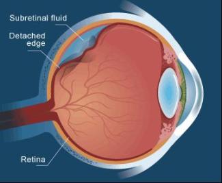
Retinal detachment is the detached retina from supporting tissue, if not treated, and then entire retina will be detached causing vision loss or blindness. It is a medical emergency.
Detachment of Retinal
Retina - is a thin light sensitive tissue layer on the eye back. The eye lens focuses light on the retina; it converts focused image into neural impulses and transmit it to the brain via the optic nerves.

The most common type of retinal detachment develops when tear/hole forms in the retina, which permits vitreous fluid from the middle of the eye to enter under the retina. This causes the retina layer to detach, so it no longer works normally and causes vision loss in the affected area.
The risk of retinal detachment is around 5 in 100,000 per year. Retinal detachment is more common among middle-aged or elderly population with rates of around 20 in 100,000 per year.
Macular degeneration
Macular degeneration is an eye condition of macula cell death (responsible for central vision), needed for reading, driving and other important tasks.
Macular degeneration
Macula - a small specialized area in the retina (the nerve layer that lines the back of the eye, senses light and creates nerve impulses that are sending through the optic nerve to the brain.) with special light sensitive cells. It helps to see fine details clearly.
Age-related macular degeneration (AMD) affects the macula, parts of the eye, which allows seeing clear images. It does not produce any pain, but it causes macula cell’s death. In some cases, AMD advances gradually so that people only notice a little change in their vision. In other cases, AMD progresses faster than usual and may lead to a vision loss in both eyes. Regular comprehensive eye exams can help diagnose macular degeneration before it causes vision loss. Proper treatment can help to slow down the vision loss, and it does not restore vision.
Macular degeneration makes it hard or even impossible to read or recognize faces, although the peripheral vision remains as usual, allow other activities of the daily life unaffected.
Cataract Surgery Complications
After cataract surgery, the eye vision improves within a few days. Vision may be blurry at first as the eye starts healing and adjusts to normality.
Recovery after Cataract Surgery
Frequent doctor’s visit is required after cataract surgery one or two days after surgery, the following week and then again, after a month to monitor healing.
It is normal to have been itching and mild discomfort for few days after surgery. Avoid rubbing or pressing on the eye. Doctor may advice to wear an eye patch or protective shield on the surgery day. Doctor may also prescribe eye drops or other medication to prevent eye infection, reduce inflammation, and control eye pressure. Complete healing occurs within eight weeks.
Contact the doctor immediately, if experience any of the following:
- Poorer vision or vision loss
- Persistent pain even after the use of over-the-counter pain medications
- Increased eye redness
- Light flashes or multiple spots in front of the eye
- Nausea, vomiting or excessive coughing
Most people rely on glasses, at least for some time after cataract surgery. Doctor will tell when the eyes have healed enough to get a final eyeglass prescription.
If have cataracts in both eyes, doctor typically schedules a second surgery a month or two later to remove the cataract in the other eye. This allows time for the first eye to heal before the second eye surgery takes place.
Cataract surgery risks & Complications
Complications after cataract surgery are uncommon, and most can treat successfully.
Cataract surgery risks include:
- Eye inflammation
- Eye infection
- Bleeding
- Swelling
- Detachment of the retina
- Glaucoma
- Secondary cataract
The risk of complications is greater if have another eye disease or other medical condition that affecting any part of the body. Occasionally, cataract surgery fails to improve vision due to the underlying eye damage from other conditions, such as glaucoma or macular degeneration.
Cataracts Surgery
Effective treatment for cataracts is surgery to remove the clouded lens of the eye. Cataract surgery is a common and safe procedure.
Cataract surgery
Cataract surgery is a procedure to remove the lens of the eye (clouded eye lens) and, in mostly replaced with an artificial lens. Cataract surgery is performing by an eye doctor (ophthalmologist) on an outpatient basis (means not have to stay in the hospital after surgery). This surgery is very common and a safe procedure.
Why does one need cataract surgery?
Cataract surgery is the best option to treat cataracts. Cataracts can cause blurry vision; if it becomes difficult to carry out the normal activities, then suggests cataract surgery.
If the cataract interferes with the treatment of another eye problem, cataract surgery may recommend. Common eye problem that may interfere with cataracts are age-related macular degeneration or diabetic retinopathy. Since cataract makes it difficult for the eye doctor to examine the back of the eye (retina & macula).
In most cases of cataract, postponing cataract surgery would not harm the eye, so have time to consider different options. If the vision is acceptably good, then may not need cataract surgery for many years. Take decision based on these questions in mind:
- How clearly can see & able to perform safely the job and to drive?
- Does you are able to read, write, see faces, or watch television?
- Does vision affects the level of independence.
- Does a bright light make you hard to see?
What happens during cataract surgery?
Cataract surgery involves removal of the clouded lens and replacing it with an artificial plastic lens. This artificial lens is installing in the same place as the natural lens and becomes part of the eye.
For some people with other eye problems stops them using an artificial lens. For them once the cataract is removing; vision can correct with eyeglasses or contact lenses.
Surgery is usually performing on one eye and after a few weeks on the other eye. Operation is performing on an outpatient basis, means not needed to stay in the hospital after surgery. During cataract surgery, eye doctor uses local anesthesia to numb the area around the eye, but the patient stay awake during the surgery. Cataract surgery is generally safe, but has the risk of infection, bleeding and increased risk of retinal detachment.
Prior to the cataract surgery
Cataract surgery takes an hour or less to perform. Eye drops placed in the eye to dilate the pupil. Local anesthetic is useful to numb the area, and may give a sedative to help the patient relax. If given a sedative, may make the patient remain awake, but groggy, during surgery.
During the cataract surgery
During surgery, the clouded natural eye lens is removing and replace with a clear artificial lens. In some cases, only clouded natural eye lens is removing without replacing it with an artificial lens.
Surgical methods used to remove cataracts include:
- Phacoemulsification – make a tiny incision in front of the eye and insert a thin needle probe, ultrasound waves are used to break (emulsify) the cataract and suck out the fragments. Lens backside is kept undisturbed and uses for the artificial lens to rest. To close the tiny incision stitches may or may not be used.
- Extracapsular extraction – make an incision in the eye to remove the lens. A less preferred procedure, because the incision is larger compared to phacoemulsification. Surgical tools are useful through this incision to remove the cloudy lens. Lens backside is kept undisturbed and uses for the artificial lens to rest. Stitching needs to close the incision.
Once the cataract has removed by either phacoemulsification or extracapsular extraction, a clear artificial lens is implanting into the empty lens capsule. This artificial lens, known as an intraocular lens (IOL), is made of plastic, acrylic, or silicone. It does not require any care and becomes a permanent part of the eye.
Varieties of IOLs available with different features
- Rigid IOL - they are rigid and implant requires several stitches to close the incision.
- Flexible IOL – they are flexible, allowing a smaller incision that requires no stitches. Surgeon can fold it, insert into the empty capsule, and then unfold inside the eye.
Some types of IOLs block ultraviolet light and others work like bifocals to provide multi-focal vision for both near and distant vision. Ask the benefits and drawbacks of the different types of IOLs with the eye surgeon to determine the best.
Cataracts Causes
Cataracts can be caused by many factors; they are age, other health conditions, eye injury and exposing to radiation or chemicals.
What causes a cataract?
- Normal aging – aging is the most common cause for cataract
- Diabetes – diabetics are at increased risk for cataract
- Smoking – increases the chances of having cataract
- Excessive alcohol consumption - believed to increase the risk of having cataract
- Eye injury or trauma – may develop a cataract immediately or after some years
- Exposure to radiation - such as ultraviolet (UV) radiation may develop a cataract
- Exposure to some harmful chemical – may develop a cataract.
- Steroidal medications – can increase the chances of cataract when used for prolong a period.
- Surgery - certain eye surgery such as glaucoma or retinal surgery may cause a cataract.
- Genetics - some time cataract from birth (inherited cataract), may be genetically caused.
Cataracts Symptoms
The most common symptom of cataracts is the noticeable blur vision and worsening of vision.
Symptoms of cataracts
- Blurred vision
- Colors appear dim and fade
- Poor night vision
- Bright lights and sunshine make glare around light
- Brown-tinted vision
- Halo around objects
- Double vision, it may decrease or disappear as the cataract grows.
If the eyesight progressively worsens, it is advisable to visit an eye specialist as soon as possible for early diagnosis and treatment.
Cataracts Types
Cataract’s types are base on its location (nuclear, cortical, and sub-capsular) and cause (Age related, secondary, traumatic, congenital, and radiation).
Cataract types based on its location
Based on their location of formation of cataract in the lens, it has three cataract types they are:
- Nuclear cataract
- Cortical cataract
- Sub-capsular cataract
Nuclear Cataracts are the most common cataract type, usually develops during aging. It affects the center or nucleus of the lens, thus it has this name. Nuclear cataract interferes with the ability to see objects, in the distance.
Cortical Cataracts are most common among diabetics. It starts forming at the outer rim or cortex of the lens and proceeds gradually toward the lens center.
Sub-capsular Cataracts develops rapidly; it affects the back of the lens or the membrane that envelops the lens. Sub-capsular cataract interferes with vision by causing glare and blurriness. It is most common among patients who use steroids, diabetics, or extreme nearsightedness.
Age is the most common risk factor for cataracts; still, some cataract types may also develop due to diabetes, eye trauma, or certain prescription drugs or steroids.
Cataract types based on its cause
Cataracts can classify into different types based on their natural of causes; they are:
- Age-related cataracts
- Secondary cataracts
- Traumatic cataracts
- Congenital cataracts
- Radiation cataracts
Age-related Cataract caused during aging, the most common type of cataract. Signs can see as early as 40 to 50 years of age, but become significant only at late 60s or 70s.
Secondary Cataracts caused by another disease or other conditions. It may develop after undergoing eye surgery, patients with diabetes or patients who are taking steroids for an extended period may develop cataracts.
Traumatic Cataract caused because of direct injury, trauma to the eye or chemical exposure. Cataracts may develop immediately or years after this triggering cause.
Congenital Cataract presents from birth, this inherited cataract in some cases is not significant enough to affect vision. If it affects vision, it should remove in order to avoid vision problems, such as strabismus or amblyopia.
Radiation Cataract caused after exposure to certain radiations (such as an ultraviolet light and other form of radiations), it is a rare form of cataract.
Cataract
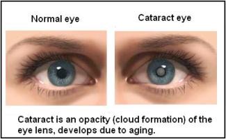
Cataract is an eye condition with cloudiness (opacity) of the eye lens that affects vision by obstructing the passage of light.
Eye cataracts
Cataracts are closely relating with the normal aging process; at an age of 80, more than half of them are effects by the cataract problem.
Eye lens – is the transparent structure behind the iris that helps to focus light rays onto the retina.
At an early stage of cataract, lens power may be increased causing myopia and slowly yellowing (opacity) of the lens causes low vision clarity. Cataract normally progresses slowly to cause vision loss or even blindness if untreated. Cataract is responsible for 48% of the world total population with blindness. Cataract usually develops in both eyes, but typically one eye progress earlier than the other does.
Eye lenses are made of water, proteins, and polysaccharides. They are arranging so precisely in a way that makes a clear lens, which allows light to pass through it and produce a clear image in the retina. Normal aging makes some of the proteins to clump together, and the area of the lens becomes cloudy. Over time the eye lens changes from transparent to cloudy yellowish brown, makes it harder to see.