Learn how chronic high blood sugar damages the blood vessels in the retina. Discover the early signs of diabetic retinopathy, screening guidelines, and how to protect your vision.
Understanding Diabetic Retinopathy
Diabetic retinopathy is a highly prevalent, progressive microvascular complication of diabetes that damages the retina, the light-sensitive tissue at the back of the eye.
Consistently elevated blood glucose levels injure the delicate microscopic blood vessels that supply the retina. If left unmanaged, these vessels can swell, leak fluid, or close off completely, cutting off vital blood flow. In advanced stages, the body grows abnormal new blood vessels that easily rupture, leading to severe vision impairment or permanent blindness.
Visual Description of Diabetic Retinopathy
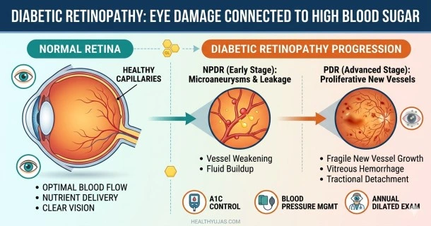
Pathologically, diabetic retinopathy manifests as structural changes inside the eye that a specialist can observe using a fundus camera or ophthalmoscope. In the early stages, the retinal background shows tiny red dots called microaneurysms, alongside small, yellowish patches of lipid deposits known as hard exudates. Fluffy white patches, called "cotton wool spots," indicate areas where nerve fibers are dying due to lack of oxygen. As the disease advances to the proliferative stage, a tangled web of fragile, abnormal new blood vessels sprouts across the retina. For the individual, this structural damage can visually manifest as a sudden shower of dark, web-like floaters, blurry patches, or dark spots that obscure fields of vision.
How Diabetes Damages Your Vision
The physiological link between hyperglycemia and retinal deterioration involves a progression of vascular changes:
- Endothelial Damage and Leakage: Chronic high blood sugar weakens the endothelial cells lining the retinal capillaries. This loss of structural integrity causes the vessel walls to balloon out (microaneurysms) and leak fluid, blood, and lipids into the surrounding retinal tissue.
- Capillary Occlusion and Ischemia: Over time, damaged capillaries become completely blocked, cutting off oxygen and nutrients to specific regions of the retina. This localized state of oxygen deprivation is known as ischemia.
- VEGF Release and Neovascularization: In response to severe ischemia, the oxygen-starved retina secretes a protein called Vascular Endothelial Growth Factor (VEGF). VEGF triggers the growth of new blood vessels (neovascularization). However, these new vessels are structurally flawed, highly fragile, and prone to leaking blood into the center of the eye.
The Core Stages of Diabetic Retinopathy
Diabetic eye disease progresses through two primary clinical classifications:
1. Nonproliferative Diabetic Retinopathy (NPDR)
This is the early stage of the disease, during which new blood vessels are not yet growing. It is divided into three sub-stages:
- Mild NPDR: The earliest stage, characterized solely by the appearance of tiny, isolated microaneurysms.
- Moderate NPDR: As the disease advances, more blood vessels swell and lose their ability to transport blood, altering the physical structure of the retina.
- Severe NPDR: A critical turning point where large numbers of blood vessels become blocked, triggering signals that force the eye to begin growing new vessels.
2. Proliferative Diabetic Retinopathy (PDR)
This is the advanced, high-risk stage of the disease. Triggered by VEGF, abnormal new blood vessels begin proliferating along the surface of the retina and into the clear, jelly-like vitreous humor that fills the eye.
- Vitreous Hemorrhage: The fragile new vessels can bleed directly into the vitreous humor, blocking light from reaching the retina and causing sudden vision loss.
- Tractional Retinal Detachment: The abnormal vessels are frequently accompanied by scar tissue. As this scar tissue shrinks, it can physically pull the retina away from the back of the eye, causing severe, permanent vision loss.
Associated Diabetes-Related Eye Conditions
In addition to retinopathy, chronic hyperglycemia increases the statistical prevalence of other serious ocular complications:
- Diabetic Macular Edema (DME): This condition occurs when leaking capillaries cause fluid buildup and swelling in the macula—the sharp, central part of the retina responsible for straight-ahead vision. DME can happen at any stage of NPDR or PDR and is the most common cause of vision loss in people with diabetes.
- Neovascular Glaucoma: If abnormal new blood vessels grow into the iris, they can block the natural drainage pathways of the eye. This blockage causes fluid pressure to build up rapidly, damaging the optic nerve.
- Accelerated Cataracts: Individuals with diabetes are statistically more likely to develop cataracts (clouding of the eye's natural lens) at a much earlier age due to high sugar concentrations altering the lens structure.
Treatment and Management Options: 3 Distinct Pathways
While advanced structural damage to the retina cannot be completely undone, multiple medical options can halt further progression and preserve remaining vision:
1. Advanced Systemic and Glycemic Control
Stabilizing daily blood glucose levels and maintaining your HbA1C within your targeted clinical range reduces the ongoing microvascular stress that drives capillary leakage. Managing blood pressure and cholesterol levels is equally vital, as hypertension directly compounds the physical pressure exerted on fragile retinal vessels.
2. Targeted Injections and Laser Therapies
Ophthalmologists can utilize precise, localized treatments to protect retinal tissue:
- Anti-VEGF Injections: Medications like aflibercept, ranibizumab, or bevacizumab are injected directly into the eye to block the effects of VEGF. This treatment reverses abnormal vessel growth and reduces swelling from macular edema.
- Panretinal Photocoagulation (PRP): A specialized laser treatment that applies tiny burns to areas of the ischemic retina away from the macula. This causes the abnormal, leaking blood vessels to shrink and fade.
- Focal Laser Therapy: Applied directly to leaking vessels in the macula to slow the leakage of fluid and stabilize vision.
3. Surgical Intervention (Vitrectomy)
For individuals with advanced Proliferative Diabetic Retinopathy, an eye surgeon can perform a vitrectomy. During this microscopic surgical procedure, the surgeon removes blood-clouded vitreous humor and clears away pulling scar tissue to prevent or repair a tractional retinal detachment.
Frequently Asked Questions (FAQ)
Can you have diabetic retinopathy without knowing it?
Yes. In its early and moderate nonproliferative stages, diabetic retinopathy rarely causes noticeable changes to your vision or physical pain. Significant structural damage and fluid leakage can occur long before your eyesight begins to blur, which is why annual comprehensive eye exams are vital.
Can diabetic retinopathy be reversed?
Early-stage nonproliferative retinopathy and macular edema can often be stabilized, managed, or partially reversed with intensive blood sugar control and targeted anti-VEGF injections. However, advanced structural changes—such as extensive retinal scarring or tissue detachment—cannot be completely reversed, though treatment can still prevent further vision loss.
How does a diabetic eye exam differ from a regular vision check?
A regular vision exam primarily measures your ability to see clearly at a distance to prescribe glasses. A comprehensive diabetic eye exam requires the use of dilating eye drops to widen your pupils. This dilation allows an ophthalmologist or optometrist to physically examine the entire retina, optic nerve, and blood vessels for microscopic signs of damage.
How often should a person with diabetes get an eye exam?
Clinical guidelines recommend that individuals with type 2 diabetes receive a comprehensive, dilated eye exam immediately upon diagnosis and at least once every year afterward. Individuals with type 1 diabetes should schedule their first dilated eye exam within five years of diagnosis and annually thereafter.