
Explore the underlying biological causes of presbyopia. Learn how changes to the eye's crystalline lens and ciliary muscles lead to blurry near vision after 40.
What Causes Presbyopia? The Anatomy and Biology of the Aging Eye
Presbyopia is an inevitable, age-related optical shift characterized by the gradual loss of your eye's ability to focus on close-up objects. To truly understand why this happens, it is important to recognize one foundational fact: presbyopia is not a disease, an illness, or an structural defect.
Instead, it is a universal, natural physiological evolution of the eye's internal structures. While refractive errors like myopia (nearsightedness), hyperopia (farsightedness), and astigmatism are typically caused by genetics or the physical shape and length of the eyeball, presbyopia is strictly caused by the natural aging process. It eventually impacts 100% of the global population, typically manifesting shortly after age 40.
The Root Biological Causes of Presbyopia
As the body grows older, the structural tissues inside the eye undergo distinct cellular and physical alterations. Presbyopia is caused by a combination of two primary internal factors operating within the eye's refractive system:
1. Progressive Hardening of the Crystalline Lens
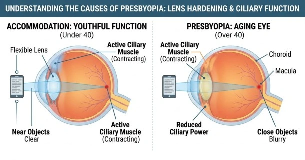
Deep inside the eye, directly behind the iris and pupil, sits the crystalline lens. In your youth, this lens is soft, clear, elastic, and highly flexible. It acts much like a flexible water balloon, easily changing its shape to modify its optical power.
Over decades of life, two structural changes take place within the lens:
- Cellular Compacting: The eye continually produces new lens fibers throughout life. Because the outer dimensions of the lens cannot expand indefinitely, these older fibers become tightly compressed and packed together.
- Protein Alterations: The natural proteins (crystallins) within the lens gradually change their structure, causing the lens matrix to become increasingly rigid.
Consequently, the crystalline lens loses its elasticity and hardens. When you attempt to look at something near, the stiffened lens can no longer bend, thicken, or increase its curvature enough to properly refract incoming light rays.
2. Changes in Ciliary Muscle Function
The lens does not change shape on its own; it relies on a ring of smooth muscle tissue called the ciliary muscle, which connects to the lens via delicate, hair-like strands called zonules.
When focusing on distant horizons, the ciliary muscles relax, pulling the zonules taut and flattening the lens. To view close-up objects, the ciliary muscles must contract, which slacks the zonules and allows a youthful, elastic lens to naturally spring into a thicker, more rounded shape.
As a normal component of aging, these microscopic ciliary muscles gradually experience a reduction in functional power. Because the ciliary muscles lack their youthful contracting force and must push against an increasingly rigid crystalline lens, the eye's entire mechanical focus system breaks down.
The Result: Loss of Eye Accommodation
The eye's natural capacity to alter its refractive power and transition focus between distant and close targets seamlessly is known as eye accommodation.
Because of lens hardening and ciliary muscle aging, the amplitude of accommodation continuously declines throughout adulthood. When light rays travel through a rigid presbyopic lens, the eye cannot bend the rays sharply enough. Instead of converging perfectly on the light-sensitive retina at the back of the eye, the focus point falls behind the retina. This anatomical displacement is what causes close-up print, text messages, and screens to look completely blurred.
- To see how these changes alter your daily life, read our detailed guide on Presbyopia Signs and Symptoms.
- Ready to explore your correction options? Visit our comprehensive overview of Presbyopia Treatments.
Secondary Risk Factors That Accelerate Onset
While normal aging is the primary cause, certain underlying medical and environmental variables can trigger premature or accelerated presbyopia (onset before the age of 40):
- Chronic Medical Conditions: Systemic illnesses such as diabetes, cardiovascular diseases, or multiple sclerosis can impair blood flow and nerve function to the eyes, accelerating structural aging.
- Pharmaceutical Medications: Regularly taking certain systemic drugs—including antihistamines, diuretics, antidepressants, and antipsychotics—can temporarily reduce the eye's natural accommodative flexibility.
- Prior Ocular Trauma: Previous eye injuries, severe intraocular inflammation, or past surgical procedures can cause localized damage to the lens or ciliary apparatus.
Frequently Asked Questions (FAQ)
Can eye exercises prevent or reverse the hardening of the crystalline lens?
No. Eye exercises cannot prevent or reverse presbyopia because they target the external extraocular muscles responsible for moving the eyeball left, right, up, and down. Eye exercises have absolutely no biochemical impact on the internal hardening of the crystalline lens or the changes occurring within the microscopic ciliary muscles.
Does having a high history of reading or close-up work cause presbyopia?
No. Engaging in extensive reading, sewing, or computer screen work will not cause or accelerate presbyopia. Intensive near-point work can trigger temporary symptoms like digital eyestrain and dry eyes, but it does not influence the underlying, biological aging rate of the internal crystalline lens matrix.
Why does presbyopia seem to happen so suddenly around age 40 if it is a gradual process?
The hardening of the crystalline lens actually begins silently in early adulthood. However, the eye possesses a massive reserve of accommodative power in youth. You only notice the condition "suddenly" around age 40 because your remaining amplitude of accommodation has finally dropped below the minimum threshold required to read comfortably at a standard working distance.